Case Reports
. 2025 Apr 26;30(1):333.
doi: 10.1186/s40001-025-02594-4.

Fully endoscopic keyhole approach for intracranial aneurysm clipping: clinical outcomes and technical note

Affiliations
Case Reports

Fully endoscopic keyhole approach for intracranial aneurysm clipping: clinical outcomes and technical note

Lulu Chen et al. Eur J Med Res. .

Abstract

Background: Surgical clipping is often utilized to treat intracranial aneurysms. The application of the endoscopy and keyhole approach in neurosurgery is increasing gradually in intracranial aneurysm occlusion. The aim of this study is to evaluate the role of fully endoscopic keyhole approach in clipping of intracranial aneurysms.

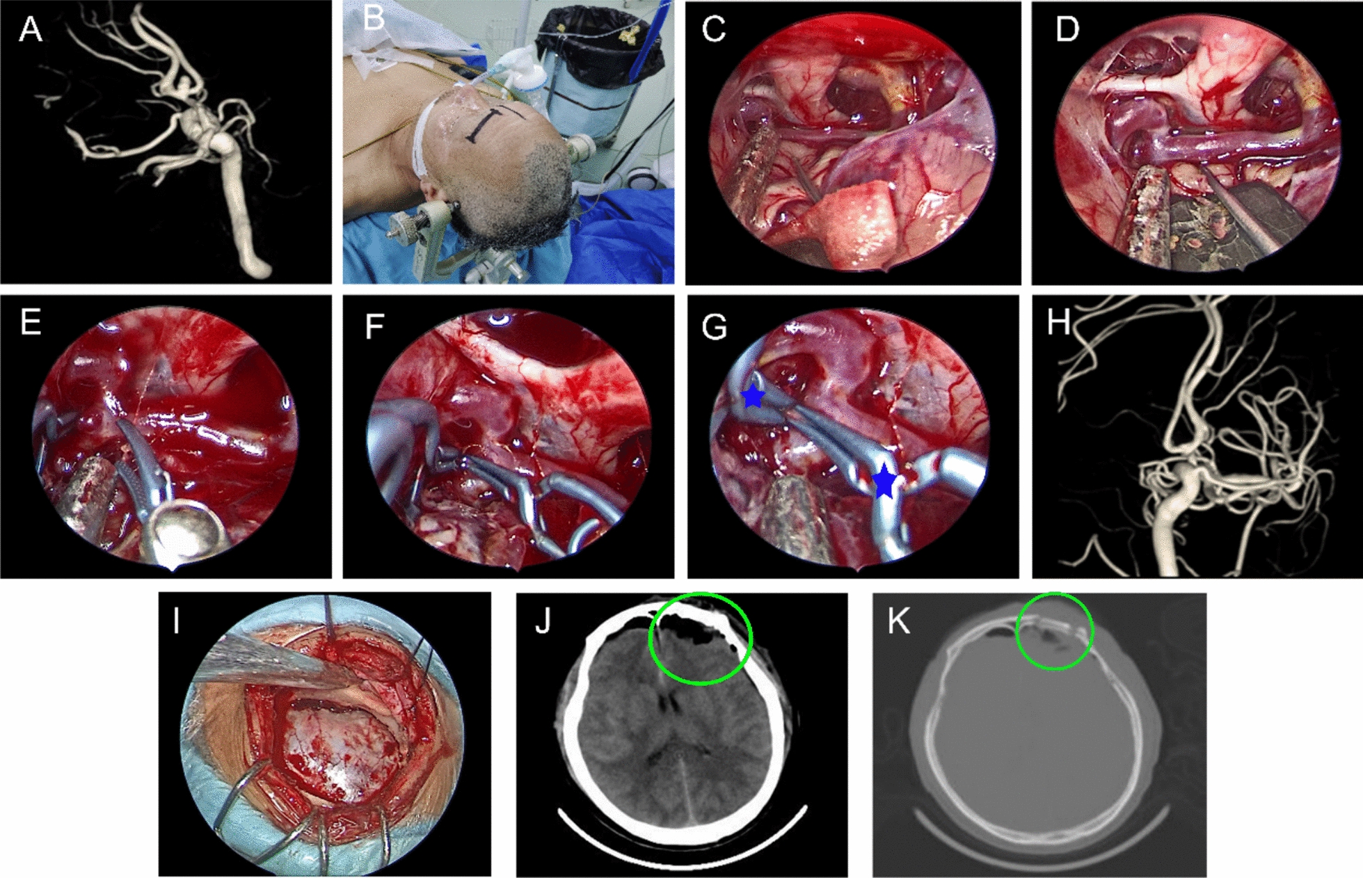
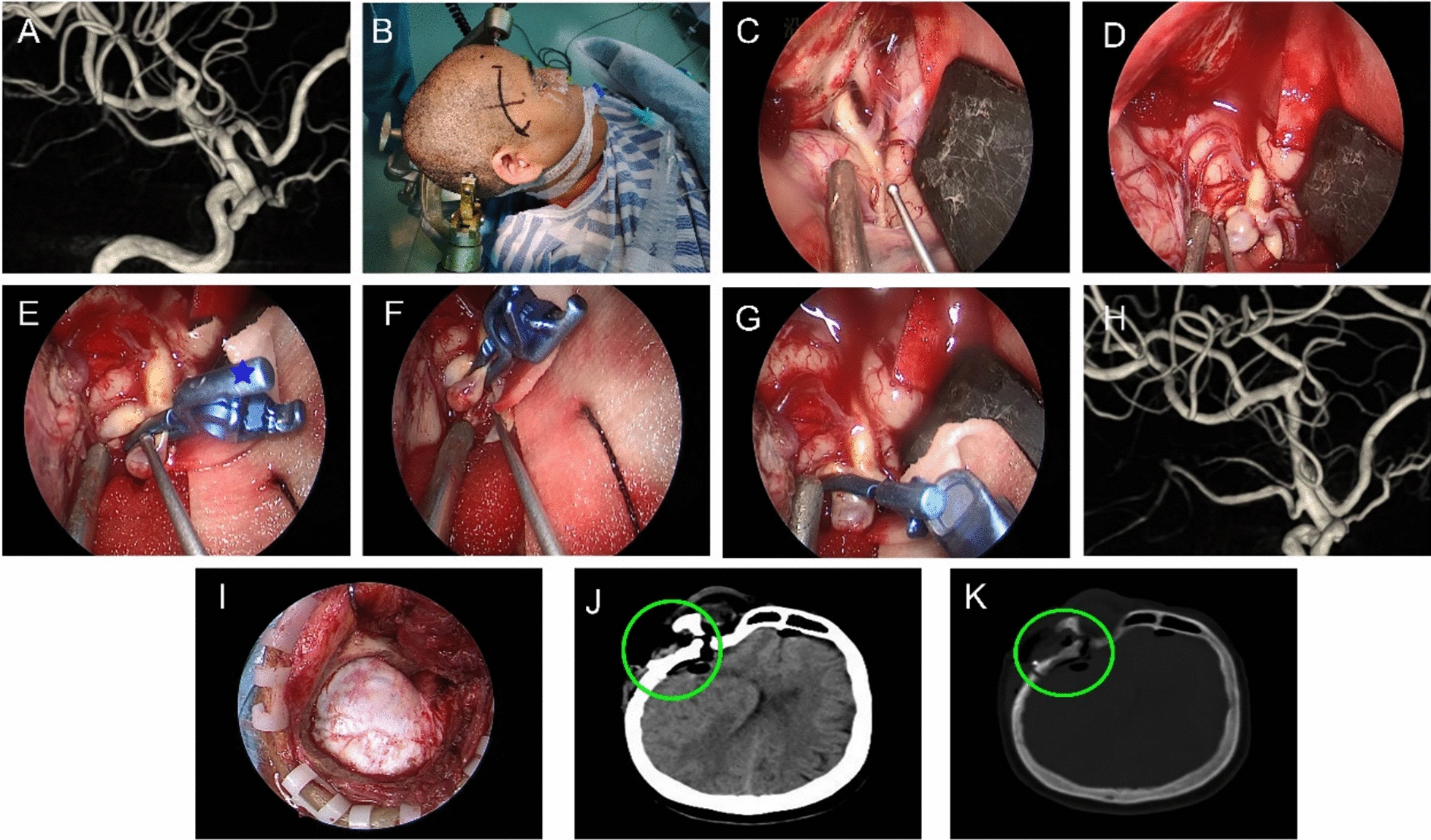
Methods: We retrospectively analyzed four cases of intracranial aneurysms, including three cases of middle cerebral artery bifurcation aneurysms (M1) and one case of anterior communicating aneurysms (ACoA). Among them, the anterior communicating aneurysm underwent fully endoscopic clipping via supraorbital keyhole approach and the middle cerebral aneurysms underwent fully endoscopic clipping via mini-pterional keyhole approach. The clipped aneurysms were evaluated by Digital Subtraction Angiography (DSA).

Results: All patients had satisfactory cerebral aneurysm clipping via the endoscopic keyhole approach. There was no cerebral hemorrhage, cerebral infarction, cerebral vasospasm. One case of intracranial infection was cured by active anti-infection. No recurrence of aneurysms after 6 months of follow-up.

Conclusion: With the advantages of the endoscopy and keyhole approach, the excellent visual field of the endoscope can reduce the influence of intracranial aneurysmal neck residual and perforating vessel. However, endoscopic clipping of intracranial aneurysms in narrow corridors requires a learning curve.

Keywords: Clipping; Endoscopy; Intracranial aneurysms; Keyhole approach; Minimally invasive.


Conflict of interest statement

Declarations. Ethics approval and consent to participate: The study was approved by the Research Ethics Committee of Bengbu Medical University. Competing interests: The authors declare no competing interests.

Figures

Fig. 1
Fig. 1
Major steps during the endoscopic eyebrow keyhole approach clipping of anterior communicating aneurysm. A Three-dimensional angiography showed the anterior communicating aneurysm. B Surgical eyebrow incision. C The carotid cistern was opened to expose the internal carotid artery and anterior cerebral artery. D Aneurysm was then dissected. EG The aneurysm was clipped and visualized with the Yasargil clip (blue five-pointed star). H Postoperative digital subtraction angiography showed a well-clipped aneurysm with no residual filling. I The extent of the bone window under the endoscopic view. J, K Postoperative CT scan indicated the extent of the craniotomy (green circle)
Fig. 2
Fig. 2
Major steps during the endoscopic mini-pterional keyhole approach clipping of a middle cerebral artery bifurcation aneurysm. A Three-dimensional angiography showed the middle cerebral artery bifurcation aneurysm. B Surgical skin incision. C The sylvian fissure and carotid cistern were opened to expose the internal carotid artery and its bifurcation. D Aneurysm was then dissected. EG The aneurysm was clipped and visualized with the Lazic aneurysm clip (blue five-pointed star). H Postoperative digital subtraction angiography showed a well-clipped aneurysm with no residual filling. I The extent of the bone window under the endoscopic view. J, K Postoperative CT scan indicated the extent of the craniotomy (green circle)

Similar articles

References

    1. Deshmukh AS, Priola SM, Katsanos AH, et al. The management of intracranial aneurysms: current trends and future directions [J]. Neurol Int. 2024;16(1):74–94. 10.3390/neurolint16010005. - PMC - PubMed
    1. Lai LT, O’Neill AH. History, evolution, and continuing innovations of intracranial aneurysm surgery [J]. World Neurosurg. 2017;102:673–81. 10.1016/j.wneu.2017.02.006. - PubMed
    1. del Maestro RF. Origin of the drake fenestrated aneurysm clip [J]. J Neurosurg. 2000;92(6):1056–64. 10.3171/jns.2000.92.6.1056. - PubMed
    1. Figueiredo EG, Deshmukh P, Nakaji P, et al. The minipterional craniotomy: technical description and anatomic assessment [J]. Neurosurgery. 2007;61(5 Suppl 2):256–64. 10.1227/01.neu.0000303978.11752.45. - PubMed
    1. Hong N, Cho WS, Pang CH, et al. Treatment outcomes of 1-stage clipping of multiple unruptured intracranial aneurysms via keyhole approaches [J]. J Neurosurg. 2022;136(2):475–84. 10.3171/2021.1.Jns204078. - PubMed

Publication types

LinkOut - more resources