. 2025 Apr 26;25(1):164.
doi: 10.1186/s12935-025-03796-w.

USP24 promotes hepatocellular carcinoma progression by deubiquitinating and stabilizing YAP1

Affiliations

USP24 promotes hepatocellular carcinoma progression by deubiquitinating and stabilizing YAP1

Huizhuang Shan et al. Cancer Cell Int. .

Abstract

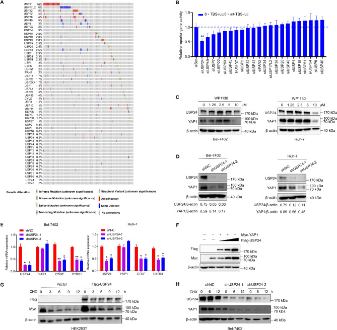
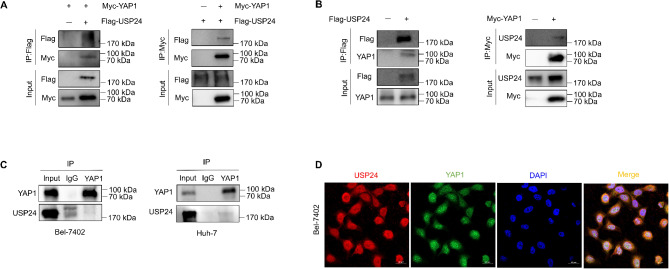
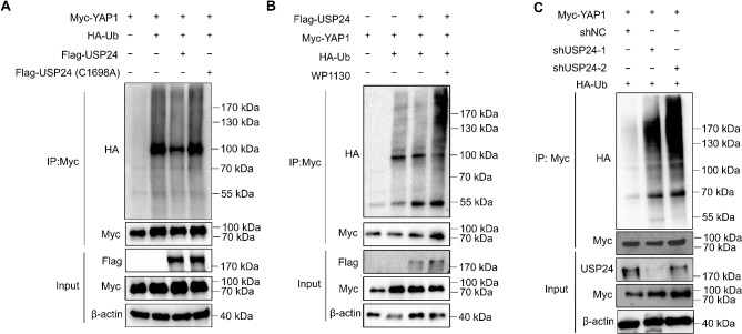
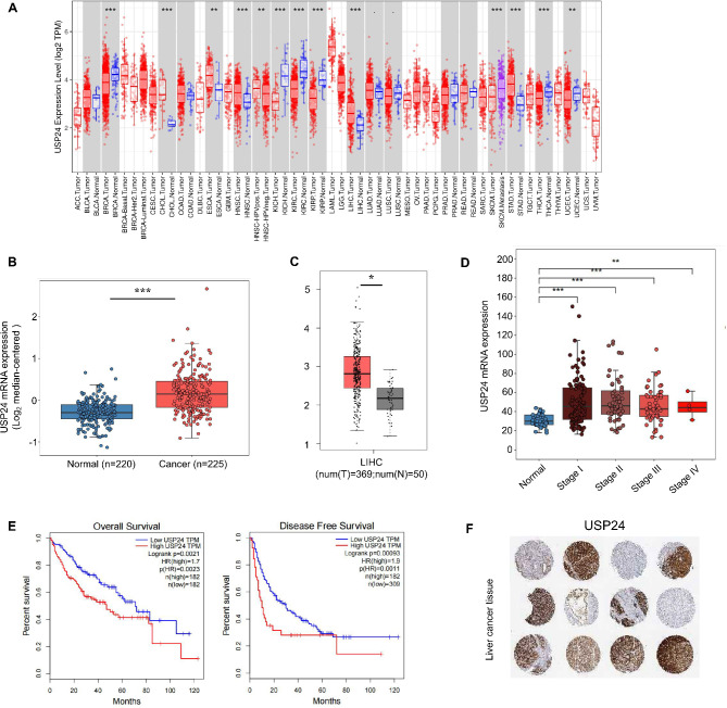
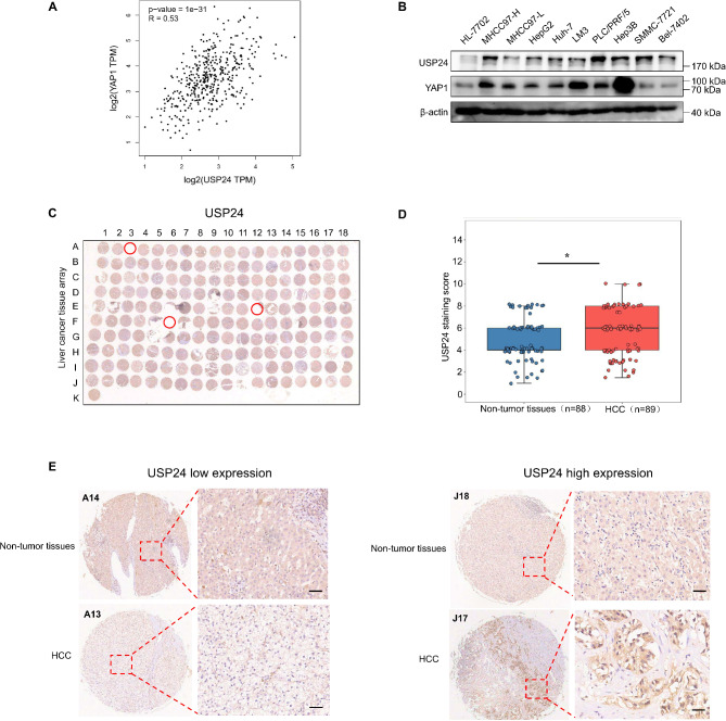
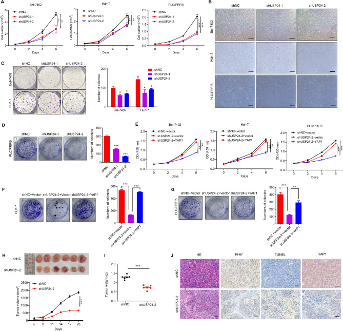
Yes-associated protein 1 (YAP1) plays a pivotal role in promoting the progression of hepatocellular carcinoma (HCC). Emerging evidence shows that inducing YAP1 degradation represents a promising strategy. Here, we identified USP24 as a bona fide deubiquitinating enzyme for YAP1. USP24 directly interacts with and deubiquitinates YAP1, thereby stabilizing YAP1 protein levels. Clinically, USP24 was significantly upregulated in HCC tissues and correlated with poor patient prognosis. Depletion of USP24 significantly suppressed the proliferation of HCC cells in vitro, which could be rescued by restoration of YAP1. Consistent with these findings, USP24 knockdown inhibited tumor growth in a xenograft mouse model. Overall, our study reveals that the USP24/YAP1 axis plays a critical role in the malignant progression of HCC, thus providing rationale for potential therapeutic interventions for YAP1-driven HCC.

Keywords: Deubiquitinating enzyme; HCC; Proliferation; USP24; YAP1.


Conflict of interest statement

Declarations. Ethics approval and consent to participate: All animal experiments were approved by The Animal Care & Welfare Committee of Guangdong Provincial People’s Hospital (Guangdong Academy of Medical Sciences), Southern Medical University. Consent for publication: Not applicable. Competing interests: The authors declare no competing interests.

Figures

Fig. 1
Fig. 1
Identification of USP24 as a regulator for YAP1 in HCC. A Analysis and generation of heat maps of CNV values of 56 USPs in the STAD dataset using cBioPortal. B After silencing USPs (CNV≥2%) in Bel-7402 cells, YAP1/TEAD4-driven transcriptional activity was accessed, and it is represented by the ratio of 8×GTIIC-luciferase activity with 8 TBSs to GTIIC luciferase activity with 8 mutant TBSs. C Bel-7402 and Huh-7 cells were treated with increasing concentrations of WP1130 for 12 h, and the indicated proteins were measured by western blotting. D USP24 was depleted by shRNAs (shUSP24-1 and shUSP24-2) in HCC cells and the indicated proteins were examined by western blotting. Control shRNA (shNC) was used as negative control. E qRT-PCR analysis of the indicated genes in Bel-7402 and Huh-7 cells with USP24 knockdown. F Increasing amounts of Flag-USP24 plasmids were co-transfected with Myc-YAP1 plasmid into HEK293T cells and the indicated proteins were examined by western blotting. G HEK293T cells were transfected with Flag-USP24 or vector plasmids. A CHX chase experiment was performed and Myc-tagged YAP1 protein was determined by western blotting (upper panel). The lower panel shows the relative protein amounts of different groups. H Bel-7402 cells stably expressing control shRNA or USP24 shRNAs were treated with or without CHX (20 mg/mL) and harvested at the indicated times, and indicated proteins was determined by western blotting (upper panel). The lower panel shows the relative protein amounts of different groups. Data are presented as mean ± SD, and P values were calculated using Student t test. *P < 0.05 and **P < 0.01
Fig. 2
Fig. 2
USP24 interacts with YAP1. A HEK293T cells were transfected with plasmids encoding Flag-USP24 and/or Myc-YAP1. After 48 h of transfection, cell extracts were prepared and immunoprecipitated with anti-Flag or anti-Myc antibodies. The protein interactions were analyzed by western blotting. B HEK293T cells were transfected with plasmids encoding Flag-USP24 or Myc-YAP1. After 48 h of transfection, cell extracts were prepared and immunoprecipitated with anti-Flag or anti-Myc antibodies, and the indicated proteins were examined by western blotting. C Whole-cell lysates from Bel-7402 or Huh-7 cells were subjected to immunoprecipitation assays with a control IgG or an anti-YAP1 antibody. The immunoprecipitates were detected by western blotting. D Confocal microscopic analysis of USP24 and YAP1 subcellular localization. Bel-7402 cells were fixed and immunostained with antibodies against the indicated proteins. Representative images from biological triplicate experiments are shown. Scale bar, 20 µM
Fig. 3
Fig. 3
USP24 deubiquitinates YAP1. A HEK293T cells were co-transfected with the specified plasmids. After 48 h of transfection, cell extracts were prepared for immunoprecipitation assays with anti-Myc followed by western blotting with anti-HA antibody. B HEK293T cells were co-transfected with the indicated plasmids, treated with or without WP1130 (10 µM) for 8 h before cells were harvested. Further, cellular extracts were prepared for immunoprecipitation assays with anti-Myc followed by western blotting with anti-HA antibody. C HEK293T cells stably expressed shRNAs specifically against USP24 and co-transfected with the indicated plasmids. Cellular extracts were immunoprecipitated with anti-Myc followed by western blotting with anti-HA antibody
Fig. 4
Fig. 4
USP24 is upregulated and associated with a poor prognosis in HCC. A USP24 mRNA expression level in different tumor tissues and non-tumor tissues from TCGA database was detected by TIMER 2.0 (http://timer.cistrome.org/). The red dots represent tumor tissues, while the blue dots represent normal tissues. B Analysis of TCGA liver cancer (Roessler Liver 2) from Oncomine database concerning the expression of USP24 in normal liver tissues and HCC tumor tissues. Data are presented by box plots. Fold change, P values (determined by Student’s t-test), and sample size are shown. C Comparison of the USP24 mRNA expression level in the TCGA-LIHC database on the GEPIA platform. D Analysis of the USP24 mRNA expression level based on the tumor stages in HCC tissues according the TCGA-LIHC database. E Prognostic survival analysis of the overall survival (OS) or disease-free survival (DFS) for USP24 gene from TCGA in HCC patients, using the GEPIA platform. The log-rank P < 0.05 is considered as a statistically significant value. The red and blue lines represent high and low expressions of USP24 gene. F Expression of USP24 in patients with HCC patients was generally high. The representative IHC images were from the Human Protein Atlas database (n = 12).*P < 0.05, **P < 0.01, and ***P < 0.001
Fig. 5
Fig. 5
USP24 is upregulated in tumor samples and correlates with the protein levels of YAP1. A Scatter plots showing the correlations between the USP24 expression level and YAP1 expression level in HCC patients by Spearman correlation analyses using GEPIA platform from TCGA database. B The USP24 and YAP1 protein level in various HCC cell lines were determined by western blotting. C Overall staining of USP24 in the tissue microarray. The microarray contained 90 pairs of HCC tissues and 90 matched normal tissues, and the detached spots (red circles) were excluded, which including 1 HCC tissue and 2 normal tissues, leaving 177 samples retained for analysis. D The USP24 staining score of HCC tissues (n = 89) and matched normal tissues (n = 88). E Representative IHC images of USP24 expression in HCC tissues and matched normal tissues. Scale bar, 50 µM. *P < 0.05
Fig. 6
Fig. 6
USP24 regulates HCC cell proliferation in vitro and in vivo.A Bel-7402, Huh-7, or PLC/PRF/5 cells were infected with control shRNA or USP24 shRNAs, and cell proliferation was monitored by trypan blue exclusion assay. B Cell morphology (96 h) was captured under the microscope. Original magnification, ×200. C Bel-7402 or Huh-7 cells were transfected with the indicated plasmids and seeded into 6-well plates at a density of 2000 cells/well. After 14 days, colony formation was detected by crystal violet staining. D PLC/PRF/5 cells were transfected with the indicated plasmids and seeded into 6-well plates at a density of 4000 cells/well. After 14 days, colony formation was detected by crystal violet staining. E HCC cells were infected with either shCtrl + vector, shUSP24-2 + vector, or shUSP24-2 + YAP1, and cell proliferation was monitored by CCK-8 assay. F, G Huh-7 (F) and PLC/PRF/5 (G) cells were infected with either shCtrl + vector, shUSP24-2 + vector, or shUSP24-2 + YAP1, and cell proliferation was monitored by colony formation assay. H, I Bel-7402 cells stably transfected with either shNC or shUSP24-2 were subcutaneously injected into BALB/c nude mice (n = 6 for each group) to establish an HCC xenograft mouse model. After 20 days, tumors from 12 of the mice were extracted and photographed. Tumor images (H, upper panel), tumor volume (H, lower panel) and weight (I) were assessed. J Expression patterns of Ki-67, TUNEL and YAP1 were examined by IHC analysis in the xenograft tumors of each group, Scale bar, 20 µM. Data are presented as mean ± SD, and P values were calculated using Student t test. *P < 0.05, **P < 0.01, and ***P < 0.001

Similar articles

References

    1. Ducreux M, Abou-Alfa GK, Bekaii-Saab T, Berlin J, Cervantes A, de Baere T, Eng C, Galle P, Gill S, Gruenberger T, et al. The management of hepatocellular carcinoma. Current expert opinion and recommendations derived from the 24th ESMO/World Congress on Gastrointestinal cancer, Barcelona, 2022. ESMO Open. 2023;8(3):101567. - PMC - PubMed
    1. Ferlay J, Ervik M, Lam F, Laversanne M, Colombet M, Mery L, Piñeros M, Znaor A, Soerjomataram I, Bray F. (2024). Global Cancer Observatory: Cancer Today. Lyon, France: International Agency for Research on Cancer. Available at https://gco.iarc.fr/today. Accessed 08-02-2024.
    1. Siegel RL, Kratzer TB, Giaquinto AN, Sung H, Jemal A. Cancer statistics, 2025. CA Cancer J Clin. 2025;75(1):10–45. - PMC - PubMed
    1. Llovet JM, Ricci S, Mazzaferro V, Hilgard P, Gane E, Blanc JF, de Oliveira AC, Santoro A, Raoul JL, Forner A, et al. Sorafenib in advanced hepatocellular carcinoma. N Engl J Med. 2008;359(4):378–90. - PubMed
    1. Llovet JM, Pena CE, Lathia CD, Shan M, Meinhardt G, Bruix J, Group SIS. Plasma biomarkers as predictors of outcome in patients with advanced hepatocellular carcinoma. Clin Cancer Res. 2012;18(8):2290–300. - PubMed

LinkOut - more resources