Review
. 2025 Apr 27;12(1):18.
doi: 10.1186/s40779-025-00603-5.

Exploring the diagnostic potential: magnetic particle imaging for brain diseases

Affiliations
Review

Exploring the diagnostic potential: magnetic particle imaging for brain diseases

Li-Shuang Guo et al. Mil Med Res. .

Abstract

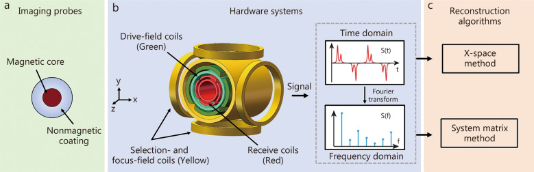
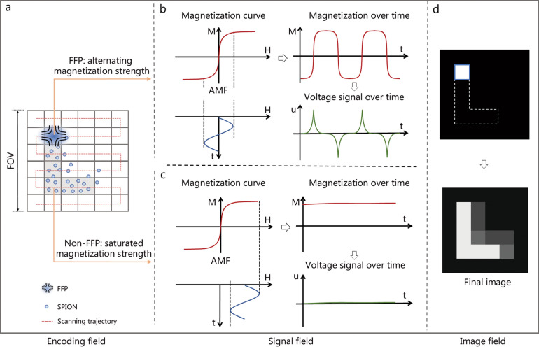
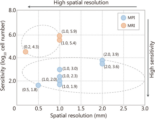
Brain diseases are characterized by high incidence, disability, and mortality rates. Their elusive nature poses a significant challenge for early diagnosis. Magnetic particle imaging (MPI) is a novel imaging technique with high sensitivity, high temporal resolution, and no ionizing radiation. It relies on the nonlinear magnetization response of superparamagnetic iron oxide nanoparticles (SPIONs), allowing visualization of the spatial concentration distribution of SPIONs in biological tissues. MPI is expected to become a mainstream technology for the early diagnosis of brain diseases, such as cancerous, cerebrovascular, neurodegenerative, and inflammatory diseases. This review provides an overview of the principles of MPI, explores its potential applications in brain diseases, and discusses the prospects for the diagnosis and management of these diseases.

Keywords: Brain diseases; Early diagnosis; Magnetic particle imaging.


Conflict of interest statement

Declarations. Ethics approval and consent to participate: Not applicable. Consent for publication: All authors consented to publish this study. Competing interests: The authors declare that they have no competing interests.

Figures

Fig. 1
Fig. 1
The component architecture diagram of magnetic particle imaging (MPI). Three key technologies for MPI are imaging probes (a), hardware systems (b), and reconstruction algorithms (c)
Fig. 2
Fig. 2
Fundamental principles of magnetic particle imaging (MPI) for field-free point (FFP). a FFP moves along a predefined trajectory in the imaging field of view (FOV). b When the FFP moves to a region containing superparamagnetic iron oxide nanoparticles (SPIONs), the SPIONs generate magnetization responses whose magnetization strength periodically changes under the drive of alternating magnetic field (AMF), and thereby producing alternating induced voltage in the receive coils. c In contrast, although driven by the same AMF as that for FFP, the SPIONs outside the FFP only generate a constant-like magnetization responses due to the magnetization saturation resulting from the selection field, and therefore producing no voltage in the receive coils. d After the FFP traverses the entire FOV, the spatial distribution of the magnetization response of SPIONs (i.e., the strength and location) is measured and recorded. After the image reconstruction, the final MPI image reflecting the spatial concentration distribution of the SPIONs is generated. M magnetization, H magnetic field, u voltage, t time
Fig. 3
Fig. 3
The development history of magnetic particle imaging (MPI) and its applications in brain diseases. After its invention in 2001, Gleich et al. [16] built the first small animal MPI system in 2005. Subsequently, several research groups conducted the exploration of MPI with different shapes [51, 52], and upgraded MPI to multimodal systems, such as MPI-magnetic resonance imaging (MRI) [53] and MPI-computed tomography (CT) systems [54]. Two commercial MPI systems were introduced in 2014 and 2016, subsequently experiencing widespread adoption. At the same time, MPI began to be applied to neuroimaging [33], where its efficacy was investigated across various diseases. With the emergence of human-sized systems [55, 56], the application scenario has been further extended to the simulation of brain diseases in human models [56]. In 2018, the concept of functional magnetic particle imaging (fMPI) was proposed [57], effectively promoting the exploration of MPI in the field of brain function [58, 59]. After 2018, the application of MPI in brain diseases has been expanded, including brain tumors [48, 65, 66], cerebrovascular diseases [60, 61, 64], and neurodegenerative diseases [62]. In 2024, the first human-scale MPI system with superconductor-based selection coils was developed [63]. FFL field-free line, FFP field-free point, CBV cerebral blood volume, SPIONs superparamagnetic iron oxide nanoparticles
Fig. 4
Fig. 4
Visual comparison of sensitivity and spatial resolution between magnetic particle imaging (MPI) and magnetic resonance imaging (MRI). The circles represent the sensitivity (vertical axis) and spatial resolution (horizontal axis), which were measured by the same MPI (blue) or the same MRI (orange). Here, the sensitivity is defined as the logarithm of the number of detectable cells. The sensitivity of MPI is within 10,000 cells [20, 30, 33, 66, 69, 70] with the highest sensitivity approximating 69 mesenchymal stem cells [68]. The sensitivities of MPI are higher than those of MRI. However, MPI slightly falls short of MRI in spatial resolution [30, 60, 68]. The metrics of MPI and MRI were measured at the small animal aperture
Fig. 5
Fig. 5
Magnetic particle imaging (MPI) detection and image-guided magnetothermal therapy for brain tumors. Imaging of orthotopic brain tumor xenografts in mice with multimodality nanoparticles (ac). In the three-dimension (3D) computerized tomography (CT)/MPI images (a) and two-dimension (2D) axial image (b) of the mouse head, the brain areas showed enhanced MPI contrast after injection compared with that of pre-injection (c). Reproduced with permission from Ref. [26] (ac). d MPI used SPIONs and an alternating magnetic field (AMF) to achieve magnetic hyperthermia for subcutaneous tumors. The heating site was controlled by moving the field-free line (FFL) to avoid thermal damage to the liver. e The top and bottom tumors in a mouse could be heated separately by moving the FFL. Reproduced with permission from Ref. [65] (d and e)
Fig. 6
Fig. 6
The potential of magnetic particle imaging (MPI) in cerebrovascular and neurodegenerative diseases. MPI was used to detect cerebral perfusion and stroke (a and b). a MPI clearly detected the ischemic area (red hash mark). b Concentration–time curves showed that MPI yields similar results to magnetic resonance imaging (MRI) but with higher temporal resolution. Reprinted with permission from Ref. [60] (a and b). The magnetoelectric effect of magnetoelectric nanoparticles (MENs) was proposed to simulate local nerve electrical activity (c and d). When coupled with MPI, it can be used to map the electric field activity of the brain in real time. c Normalized de-modulated MPI-MEN images of 2 different views of frontal lobes. d The insert shows a detailed normalized three-dimensional field profile in the region of firing. Reproduced with permission from Ref. [59] (c and d)
Fig. 7
Fig. 7
The human-size magnetic particle imaging (MPI) for stroke. a In static stroke experiments, concentration differences can be observed in the stroke parts compared to control measurements. b The complete phantom, consisting of two tubes filled with glass spheres, was used to simulate blood flow in vessels. c Dynamic imaging results of the brain imager, including time to peak (TTP), mean transit time (MTT), relative cerebral blood flow (rCBF) and relative cerebral blood volume (rCBV). Reprinted with permission from Ref. [56] (ac). FOV field of view

Similar articles

Cited by

References

    1. Liu H, Zhang J, Chen X, Du XS, Zhang JL, Liu G, et al. Application of iron oxide nanoparticles in glioma imaging and therapy: from bench to bedside. Nanoscale. 2016;8(15):7808–26. - PubMed
    1. Sweeney MD, Zhao Z, Montagne A, Nelson AR, Zlokovic BV. Blood-brain barrier: from physiology to disease and back. Physiol Rev. 2019;99(1):21–78. - PMC - PubMed
    1. Jiang J, Gao G, Feng J, Mao Q, Chen L, Yang X, et al. Traumatic brain injury in China. Lancet Neurol. 2019;18(3):286–95. - PubMed
    1. Campbell BCV, Khatri P. Stroke. Lancet. 2020;396(10244):129–42. - PubMed
    1. Wardlaw JM, Smith C, Dichgans M. Mechanisms of sporadic cerebral small vessel disease: insights from neuroimaging. Lancet Neurol. 2013;12(5):483–97. - PMC - PubMed

MeSH terms

Substances

LinkOut - more resources