

Auto-segmentation, radiomic reproducibility, and comparison of radiomics between manual and AI-derived segmentations for coronary arteries in cardiac [18F]NaF PET/CT images
- PMID: 40287890
- PMCID: PMC12034606
- DOI: 10.1186/s40658-025-00751-6
Auto-segmentation, radiomic reproducibility, and comparison of radiomics between manual and AI-derived segmentations for coronary arteries in cardiac [18F]NaF PET/CT images
Abstract
Background: [18F]NaF is a potential biomarker for assessing cardiac risk. Automated analysis of [18F]NaF positron emission tomography (PET) images, specifically through quantitative image analysis ("radiomics"), can potentially enhance diagnostic accuracy and personalised patient management. However, it is essential to evaluate the reproducibility and reliability of radiomic features to ensure their clinical applicability. This study aimed to (i) develop and evaluate an automated model for coronary artery segmentation using [18F]NaF PET and calcium scoring computed tomography (CSCT) images, (ii) assess inter- and intra-observer radiomic reproducibility from manual segmentations, and (iii) evaluate the radiomics reliability from AI-derived segmentations by comparison with manual segmentations.
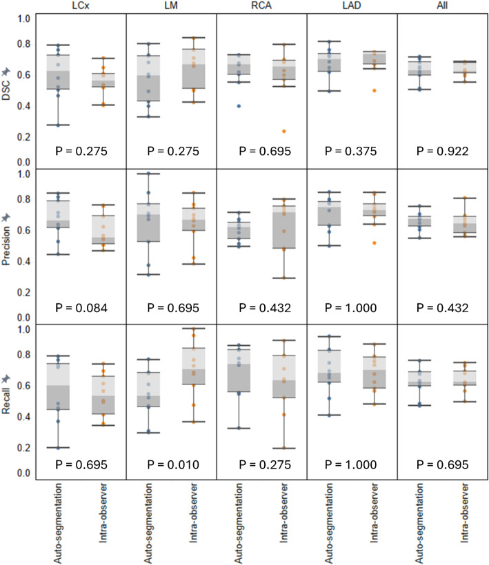
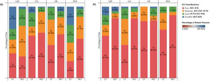
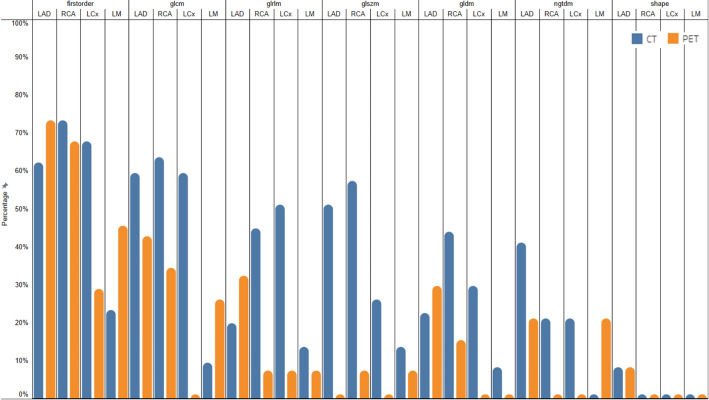
Results: 141 patients from the "effects of Vitamin K and Colchicine on vascular calcification activity" (VikCoVac, ACTRN12616000024448) trial were included. 113 were used to train an auto-segmentation model using nnUNet on [18F]NaF PET and CSCT images. Reproducibility of inter- and intra-observer radiomics and reliability of radiomics from AI-derived segmentations was assessed using lower bound of intraclass correlation coefficient (ICC). The auto-segmentation model achieved an average Dice Similarity Coefficient of 0.61 ± 0.05, having no statistically significant difference compared to the intra-observer variability (p = 0.922). For the unfiltered images, 47(12.6%) CT and 25(7.5%) PET radiomics were inter-observer reproducible, while 133(35.8%) CT and 57(15.3%) PET radiomics were intra-observer reproducible. 7(9.7%) CT and 18(25.0%) PET first-order features, as well as 17(17.7%) CT GLCM features, were reproducible for both inter- and intra-observer analyses. 9.8% and 16.8% of radiomics from AI-derived segmentations showed excellent and good reliability. First-order features were most reliable (ICC > 0.75; 78/144[54.2%]) and shape features least (2/112[1.8%]). CT features demonstrated greater reliability (147/428[34.3%]) than PET (81/428 [18.9%]). Features from the left anterior descending (76/214[35.5%]) and right coronary artery (75/214[35.0%]) were more reliability than the circumflex (49/214[22.9%]) and left main (28/214[13.1%]) arteries.
Conclusions: An effective segmentation model for coronary arteries was developed and reproducible [18F]NaF PET/CSCT radiomics were identified through inter- and intra-observer assessments, supporting their clinical applicability. The reliability of radiomics from AI-derived segmentations compared to manual segmentations was highlighted. The novelty of [18F]NaF as a biomarker underscores its potential in providing unique insights into vascular calcification activity and cardiac risk assessment.
Clinical trial registration: VIKCOVAC trial ("effects of Vitamin K and Colchicine on vascular calcification activity"). Unique identifier: ACTRN12616000024448. URL: https://www.anzctr.org.au/Trial/Registration/TrialReview.aspx?id=368825 .
Keywords: Auto-segmentation; Coronary artery disease; Radiomics; Reproducibility; [18F]NaF PET.
© 2025. The Author(s).
Conflict of interest statement
Declarations. Ethics approval and consent to participate: Ethics approval for undertaking this study was acquired from the Royal Perth Hospital Human Research and Ethics Committee (REG14-095) and the study was performed in accordance with the ethical standards as laid down in the 1964 Declaration of Helsinki and its later amendments or comparable ethical standards. All participants provided written informed consent. Consent for publication: All participants provided written informed consent. Competing interests: The authors declare that they have no competing interests.
Figures


Similar articles
-
A Critical Analysis of the Robustness of Radiomics to Variations in Segmentation Methods in 18F-PSMA-1007 PET Images of Patients Affected by Prostate Cancer.Diagnostics (Basel). 2023 Dec 11;13(24):3640. doi: 10.3390/diagnostics13243640. Diagnostics (Basel). 2023. PMID: 38132224 Free PMC article.
-
Reproducibility of F18-FDG PET radiomic features for different cervical tumor segmentation methods, gray-level discretization, and reconstruction algorithms.J Appl Clin Med Phys. 2017 Nov;18(6):32-48. doi: 10.1002/acm2.12170. Epub 2017 Sep 11. J Appl Clin Med Phys. 2017. PMID: 28891217 Free PMC article.
-
Automated multiclass segmentation, quantification, and visualization of the diseased aorta on hybrid PET/CT-SEQUOIA.Med Phys. 2024 Jun;51(6):4297-4310. doi: 10.1002/mp.16967. Epub 2024 Feb 7. Med Phys. 2024. PMID: 38323867
-
A Scoping Review of Machine-Learning Derived Radiomic Analysis of CT and PET Imaging to Investigate Atherosclerotic Cardiovascular Disease.Tomography. 2024 Sep 3;10(9):1455-1487. doi: 10.3390/tomography10090108. Tomography. 2024. PMID: 39330754 Free PMC article.
-
Current progress and quality of radiomic studies for predicting EGFR mutation in patients with non-small cell lung cancer using PET/CT images: a systematic review.Br J Radiol. 2021 Jun 1;94(1122):20201272. doi: 10.1259/bjr.20201272. Epub 2021 May 12. Br J Radiol. 2021. PMID: 33882244 Free PMC article.
References
-
- Martin SS, et al. 2024 heart disease and stroke statistics: A report of US and global data from the American heart association. Circulation. 2024;149(8):e347–913. - PubMed
-
- Budoff MJ, et al. Progression of coronary artery calcium predicts All-Cause mortality. Jacc-Cardiovasc Imag. 2010;3(12):1229–36. - PubMed
-
- Budoff MJ, et al. Assessment of coronary artery disease by cardiac computed tomography - A scientific statement from the American heart association committee on cardiovascular imaging and intervention, Council on cardiovascular radiology and intervention, and committee on cardiac imaging, Council on clinical cardiology. Circulation. 2006;114(16):1761–91. - PubMed
Grants and funding
LinkOut - more resources
Full Text Sources
Medical
