Review
. 2025 Apr 28;26(1):89.
doi: 10.1186/s10194-025-01978-5.

Beyond the "string of beads": case-based exploration of diagnostic pitfalls and solutions in reversible cerebral vasoconstriction syndrome

Affiliations
Review

Beyond the "string of beads": case-based exploration of diagnostic pitfalls and solutions in reversible cerebral vasoconstriction syndrome

Seung Ae Kim et al. J Headache Pain. .

Abstract

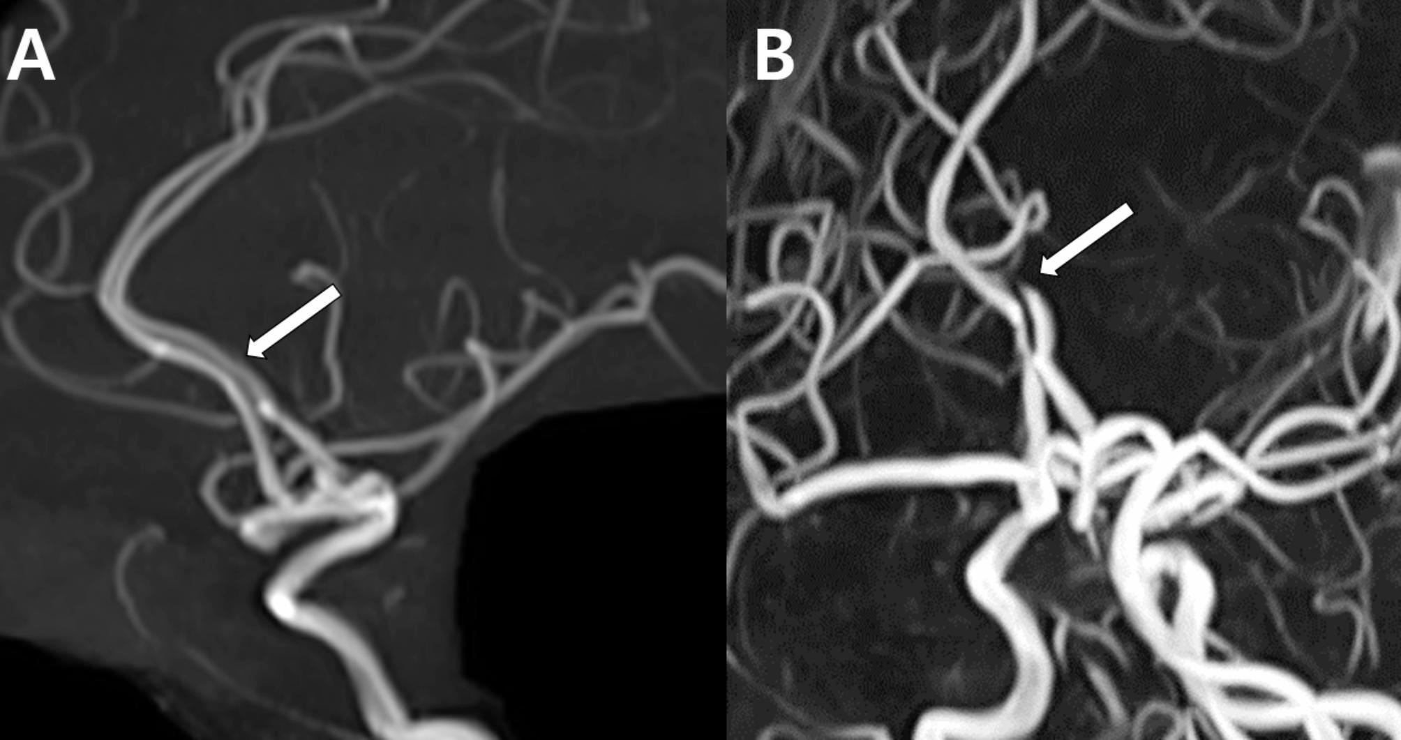
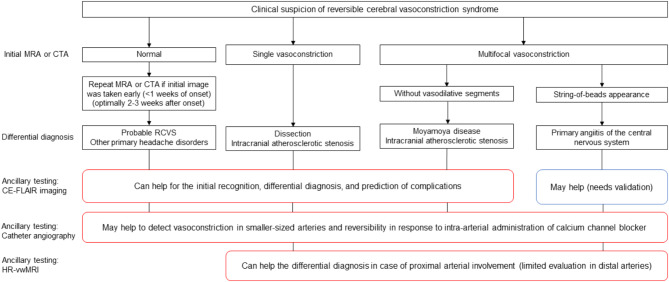
Background: The diagnosis of reversible cerebral vasoconstriction syndrome (RCVS) is challenging due to its varied clinical manifestations and imaging findings. While it typically presents with a sudden, severe thunderclap headache and multifocal constriction of the cerebral arteries, the wide spectrum of radiological presentations may complicate the diagnosis.

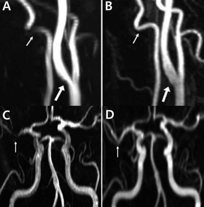
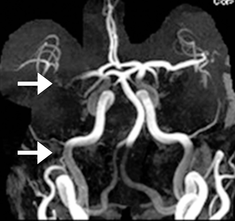
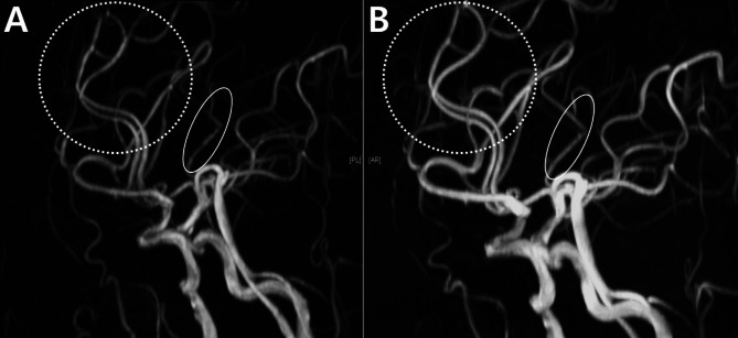
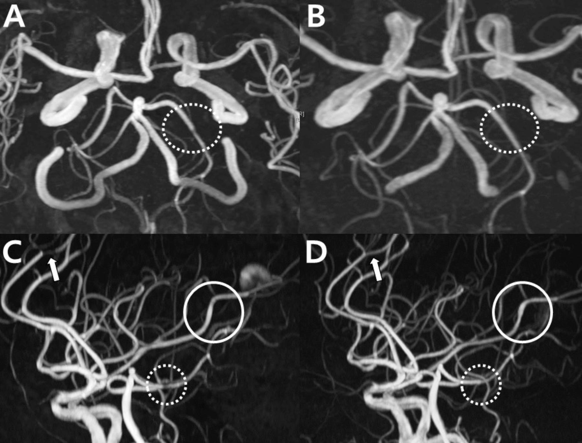
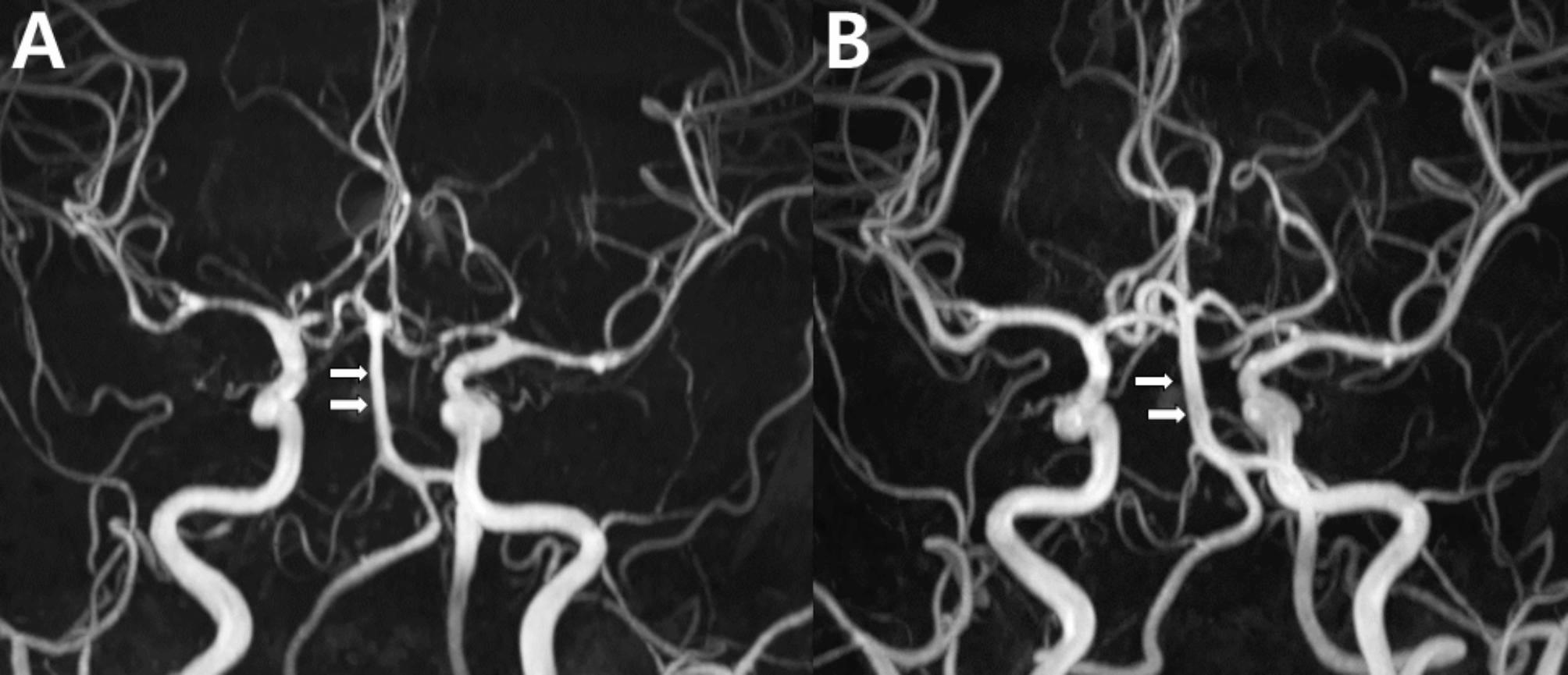
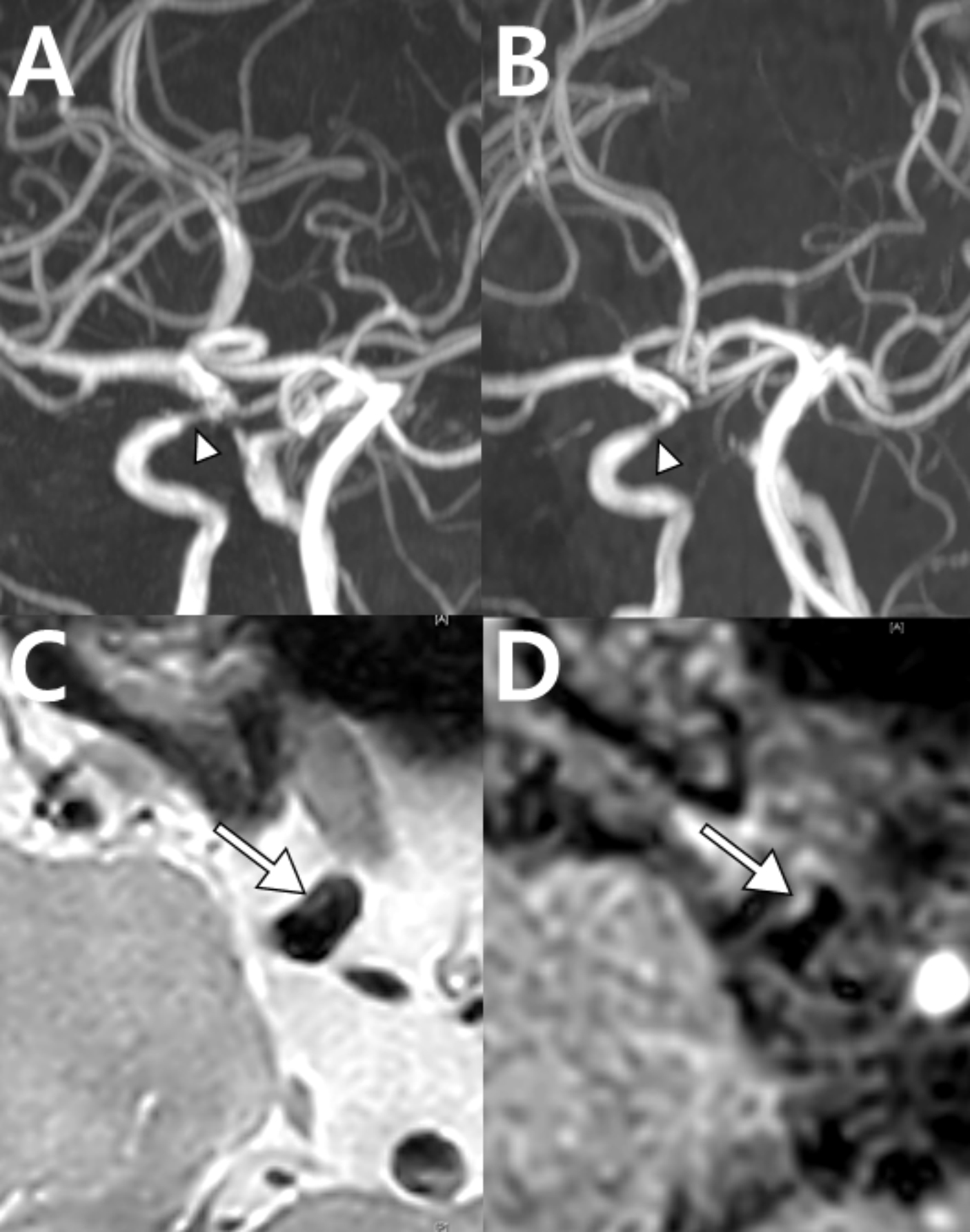
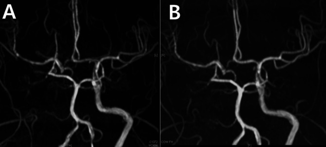
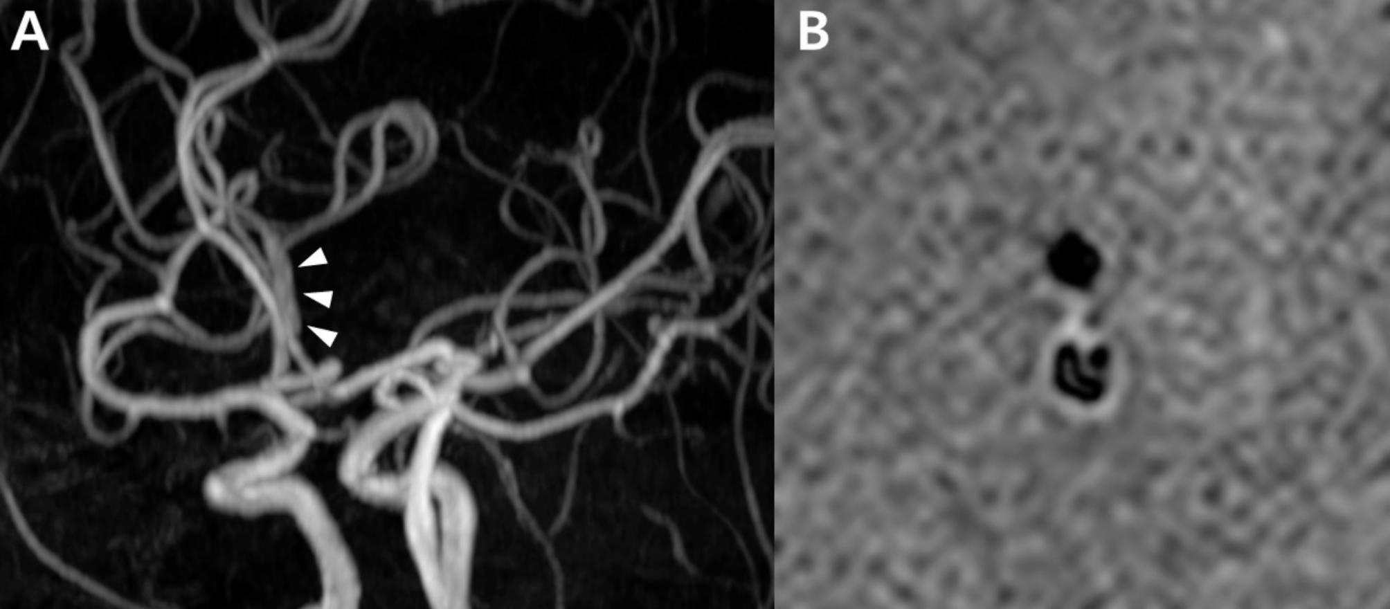
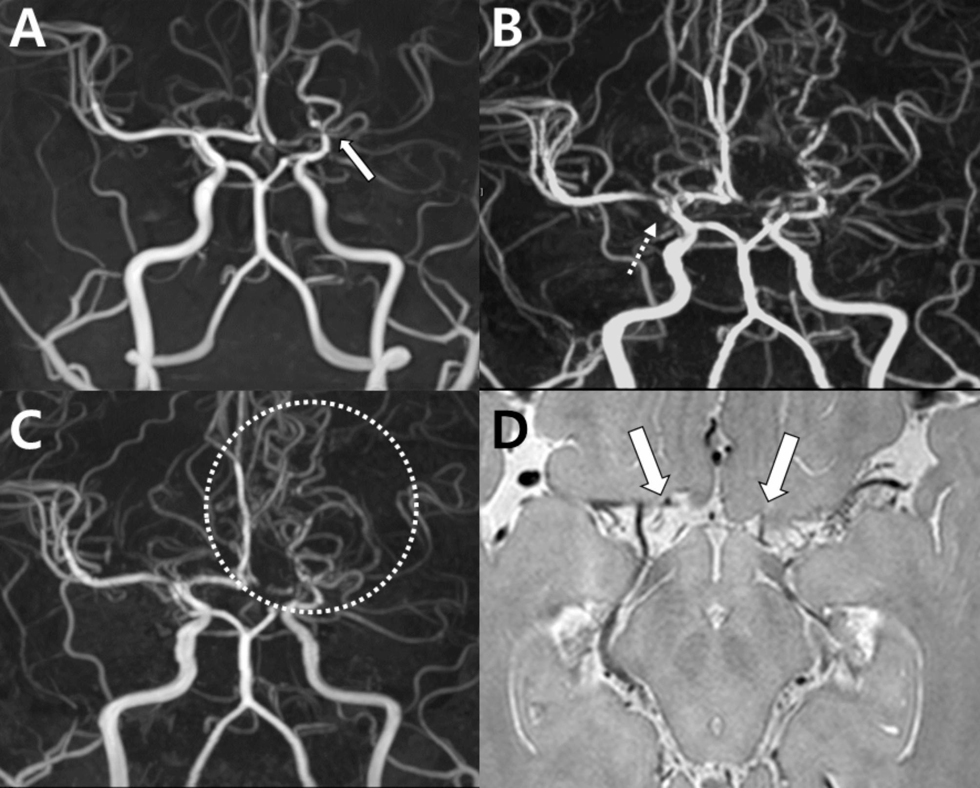
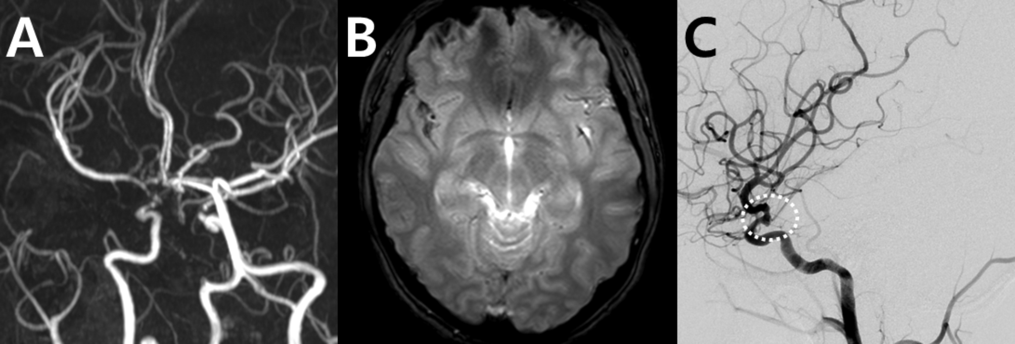
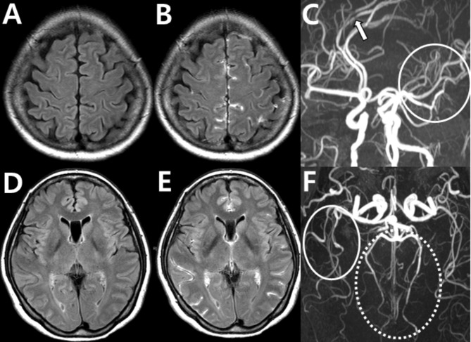
Main body: This review presents a series of cases that show both typical and atypical presentations of RCVS. Typical cases show the characteristic "string of beads" pattern on angiography, which usually resolves within 3-6 months. However, diagnostic challenges arise when angiography appears normal in the early stages or when imaging artifacts obscure the findings. In addition, the variability in vasoconstriction patterns and the need for a differential diagnosis further complicate the accurate identification. These cases highlight the importance of considering RCVS in patients with recurrent thunderclap headaches, even when the initial imaging is inconclusive. Recognizing these challenges and the variability in presentation, along with the use of high-resolution vessel wall MRI and blood-brain barrier imaging, can improve diagnostic accuracy and improve patient outcomes.

Conclusion: The diagnosis of RCVS requires careful integration of clinical evaluation and advanced imaging techniques, with particular attention to radiological findings that can guide accurate diagnosis and management. Despite challenges, such as normal early stage angiography and imaging variability, maintaining a high suspicion of RCVS is essential, especially in patients with recurrent thunderclap headaches.

Keywords: Angiography; Blood-brain barrier imaging; Cerebral vasoconstriction; Differential diagnosis; High-resolution vessel wall MRI; Imaging artifacts; Reversible cerebral vasoconstriction syndrome; Thunderclap headache.


Conflict of interest statement

Declarations. Ethics approval and consent to participate: Not applicable. Consent for publication: Not applicable. Competing interests: The authors declare no competing interests.

Figures

Fig. 1
Fig. 1
Typical steno-dilatation in reversible cerebral vasoconstriction syndrome: case 1. Time-of-flight magnetic resonance angiography (TOF MRA) image taken 17 days after onset in a pregnant woman with RCVS showed segmental stenosis (arrowheads: highlighting only selected regions for clarity) in multiple cerebral arteries and post-stenotic dilatation in the basilar artery (arrow) and other cerebral arteries, presenting the characteristic “string of beads” or “sausage-on-a-string” pattern of RCVS
Fig. 2
Fig. 2
Typical steno-dilations in reversible cerebral vasoconstriction syndrome: case 2. Multiple segmental stenoses (arrow heads) and dilations (arrows) in the distal middle cerebral arteries (MCAs) and posterior cerebral arteries (PCAs) in a 56-year-old patient with RCVS experienced a thunderclap headache triggered by brushing teeth, coughing, bending, tying hair, and speaking in tense situations. Only representative regions are marked for clarity
Fig. 3
Fig. 3
Reversible cerebral vasoconstriction syndrome with normal brain magnetic resonance angiography findings. Brain MRA of a 31-year-old patient with recurrent thunderclap headaches triggered by typical precipitants, which did not show significant abnormalities
Fig. 4
Fig. 4
Stair-step artifacts. (A) Pseudo-stenosis caused by stair-step artifacts in 2D TOF MRA is observed in the right internal carotid artery (ICA) (thick arrow), left ICA (thin arrow), bilateral vertebral arteries, and left common carotid artery (dotted arrows). (B) In the contrast-enhanced MRA of the same patient, no artifacts are seen in the vessels indicated in (A). Figure adapted from McKinney AM: Artifacts of the Craniocervical Arterial System on MRI. In: Atlas of Normal Imaging Variations of the Brain, Skull, and Craniocervical Vasculature. Cham: Springer International Publishing; 2017: 1261–1291, under the terms of the Creative Commons Attribution License [16]
Fig. 5
Fig. 5
In-plane saturation artifact. (A) Pseudo-stenosis in the ICA (thick arrow) and in-plane saturation artifacts in the VA (thin arrow) are observed in a 2D TOF MRA. (B) The contrast-enhanced MRA shows normal findings in the previously observed areas. (C) A pseudo-stenosis that appears as an artificial defect in the right MCA (arrow) is evident on a 3D TOF MRA. (D) However, this defect is not seen in the contrast-enhanced MRA image. Figure adapted from McKinney AM: Artifacts of the Craniocervical Arterial System on MRI. In: Atlas of Normal Imaging Variations of the Brain, Skull, and Craniocervical Vasculature. Cham: Springer International Publishing; 2017: 1261–1291, under the terms of the Creative Commons Attribution License [16]
Fig. 6
Fig. 6
Venetian blind artifacts in 3D TOF imaging resulting from signal loss due to repeated radiofrequency pulses, particularly at slab edges (arrows). Figure adapted from Sayah A, Mamourian AC: Flow-Related Artifacts in MR Imaging and MR Angiography of the Central Nervous System. Neurographics 2012, 2(4):154–162, under the terms of the Creative Commons Attribution License [19]
Fig. 7
Fig. 7
Reversible cerebral vasoconstriction syndrome with ambiguous findings: case 4. (A) An initial MRA performed 6 days after onset shows uncertainty between distal artery visualization issues and possible stenosis, particularly in the anterior cerebral arteries (ACAs) (dotted circles) and the MCA (oval). (B) Follow-up MRA 3 months later reveals distal vessel dilation in the same ACAs (dotted circles) and MCA (oval), suggesting RCVS
Fig. 8
Fig. 8
Rapid reversibility of stenosis and new focal dilation in reversible cerebral vasoconstriction syndrome: Case 5. (A) Initial MRA with focal stenosis in the left PCA (dotted circle). (B) Follow-up MRA in 2 days showing normalized stenosis (dotted circle). (C) Initial MRA suspicious for stenosis vs. artifact in the right anterior (arrow) and left PCAs (dotted circle). (D) The follow-up MRA shows normalization of the right anterior (arrow) and left PCAs (dotted circle) and a focal dilation in the left MCA (circle) newly developed
Fig. 9
Fig. 9
Diffuse narrowing of the basilar artery as a reversible cerebral vasoconstriction syndrome phenomenon: case 6. (A) Multifocal narrowing of the bilateral M1, A1, and A2 segments is observed in the initial time-of-flight MR angiography of a 30-year-old patient with postpartum RCVS. In this imaging, the basilar artery (arrows) appeared normal. (B) The follow-up imaging obtained 3 months later showed normalization of the overall vessels. The diameter of the basilar artery increased compared to the initial imaging (arrows), indicating that there is a diffuse narrowing in the basilar artery, which can be missed if assessed cross-sectionally
Fig. 10
Fig. 10
Mimickers of reversible cerebral vasoconstriction syndrome: focal intracranial atherosclerosis in a patient with reversible cerebral vasoconstriction syndrome (case 7). (A) Multifocal stenosis is observed in the ACA, MCA, and most prominently in the right distal ICA (arrowhead) in the initial time-of-flight MR angiography. (B) Six months later, overall vasospasm improved, except for the right distal ICA (arrowhead). (C, D) High-resolution vessel wall MRI (C, T2 weighted image; D, T1 post-contrast image) show an atherosclerotic plaque in the right distal ICA (arrow)
Fig. 11
Fig. 11
Mimickers of reversible cerebral vasoconstriction syndrome: asymptomatic atherosclerosis (case 8). (A) MR angiography shows right ICA occlusion with multifocal stenosis in the distal M1, M2, M3, A3, and V4 segments in a 74-year-old patient with clinically suspected RCVS. (B) There is no significant change 1 year after onset. The diagnosis of asymptomatic intracranial atherosclerosis and probable RCVS (angiogram-negative) is made
Fig. 12
Fig. 12
Mimickers of reversible cerebral vasoconstriction syndrome: Intracranial arterial dissection (case 9). The MRA shows a single dilation (arrowhead) in the left A2 segment in a patient with thunderclap headache (Case 9), who is ultimately diagnosed with dissection by (B) high-resolution vessel wall MRI revealing a dissecting flap
Fig. 13
Fig. 13
Reversible cerebral vasoconstriction syndrome resembling intracranial arterial dissection (case 10). (A) MRA taken 2 days after thunderclap headache showed normal findings in the left ACA (arrow). (B) However, a single flame-shaped occlusion in the left ACA (arrow) was observed 8 days after onset, suggesting RCVS rather than arterial dissection due to its dynamic imaging characteristics
Fig. 14
Fig. 14
Mimickers of reversible cerebral vasoconstriction syndrome: unilateral moyamoya disease with rapid progression to bilateral disease (case 11). (A-C) Serial MRA angiographic imaging in a 30-year-old postpartum patient with recurrent transient ischemic attacks, right arm paralysis, and mild headache. (A) Initial imaging on the first day of onset shows isolated left distal ICA occlusion (arrow). (B) Subsequent imaging at 8 days after onset revealed rapid progression to right distal ICA stenosis (dotted arrow) along with left distal multifocal stenosis and a string-of-beads pattern. (C) At 1 year, no reversal of the bilateral distal ICA steno-occlusion or the vasoconstriction in the left distal arteries with formation of basal collateral vessels (dotted circle) were observed. (D) High-resolution vessel wall MRI showed significant obliteration of the ICAs (arrows), suggesting moyamoya disease. These cumulative observations led to the final diagnosis of moyamoya disease
Fig. 15
Fig. 15
Mimickers of reversible cerebral vasoconstriction syndrome: aneurysmal subarachnoid hemorrhage and vasospasm (case 12). A 23-year-old female who had severe headache episodes was initially misdiagnosed with RCVS. (A) Magnetic resonance angiography (MRA) shows severe stenosis in the right distal ICA and M1 segment of the ACAs. (B) A small amount of subarachnoid hemorrhage is observed along the right Sylvian fissure in the gradient-echo image. (C) Catheter angiography reveals a ruptured aneurysm over the right distal ICA aneurysm (dotted circles), requiring emergency coil embolization
Fig. 16
Fig. 16
Blood-brain barrier disruption in reversible cerebral vasoconstriction syndrome. BBB breakdown is observed using contrast-enhanced FLAIR imaging in a 56-year-old female with RCVS (case 2). (A, D) FLAIR images without contrast. (B, E) The same sections with contrast-enhanced FLAIR MRI show BBB disruption through hyperintense CSF. (C, F) MRA shows typical multifocal steno-dilatation in the ACA (arrow), left MCA (solid circles), and distal PCAs (dotted circles)
Fig. 17
Fig. 17
Diagnostic flow chart for radiologic evaluation and differential diagnosis in suspected reversible cerebral vasoconstriction syndrome. **Abbreviations: MRA, Magnetic Resonance Angiography; CTA, Computed Tomography Angiography; RCVS, Reversible Cerebral Vasoconstriction Syndrome; CE-FLAIR, Contrast-Enhanced Fluid-Attenuated Inversion Recovery; HR-vwMRI, High-Resolution Vessel Wall Magnetic Resonance Imaging;

Similar articles

References

    1. Calabrese LH, Dodick DW, Schwedt TJ, Singhal AB (2007) Narrative review: reversible cerebral vasoconstriction syndromes. Ann Intern Med 146(1):34–44 - PubMed
    1. Ducros A (2012) Reversible cerebral vasoconstriction syndrome. Lancet Neurol 11(10):906–917 - PubMed
    1. Cho SH, Kim BK, Lee MJ (2022) Clinical characteristics of reversible cerebral vasoconstriction syndrome: a large korean multicenter study. Headache Pain Res 23(2):27–32.
    1. Headache Classification Committee of the International Headache Society (IHS) (2018) The International classification of Headache disorders, 3rd edition. Cephalalgia 38(1):1–211 - PubMed
    1. Song TJ, Lee KH, Li H, Kim JY, Chang K, Kim SH, Han KH, Kim BY, Kronbichler A, Ducros A et al (2021) Reversible cerebral vasoconstriction syndrome: a comprehensive systematic review. Eur Rev Med Pharmacol Sci 25(9):3519–3529 - PubMed

LinkOut - more resources