

TiO2 Nanotube Implants Modified with Silk Fibroin and Mesoporous Silica Nanocomposite Coatings Enable Efficient Drug Release to Promote Osteogenesis
- PMID: 40289330
- PMCID: PMC12123568
- DOI: 10.1021/acsami.5c03599
TiO2 Nanotube Implants Modified with Silk Fibroin and Mesoporous Silica Nanocomposite Coatings Enable Efficient Drug Release to Promote Osteogenesis
Abstract
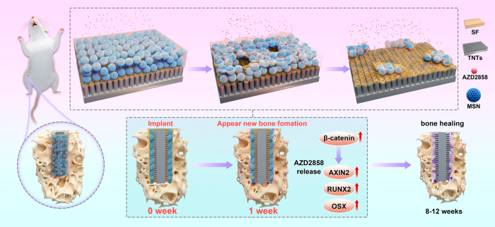
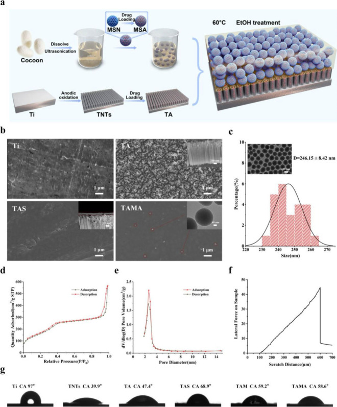
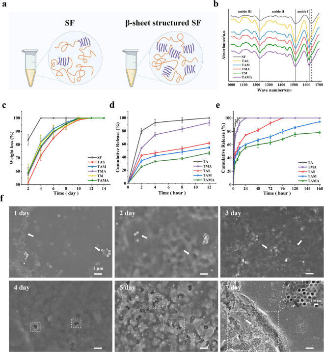
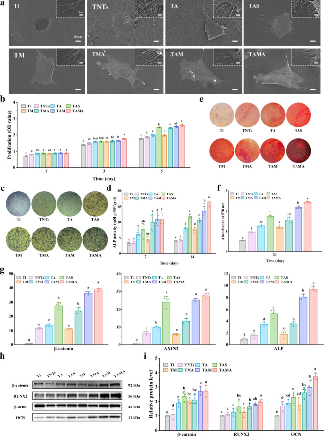
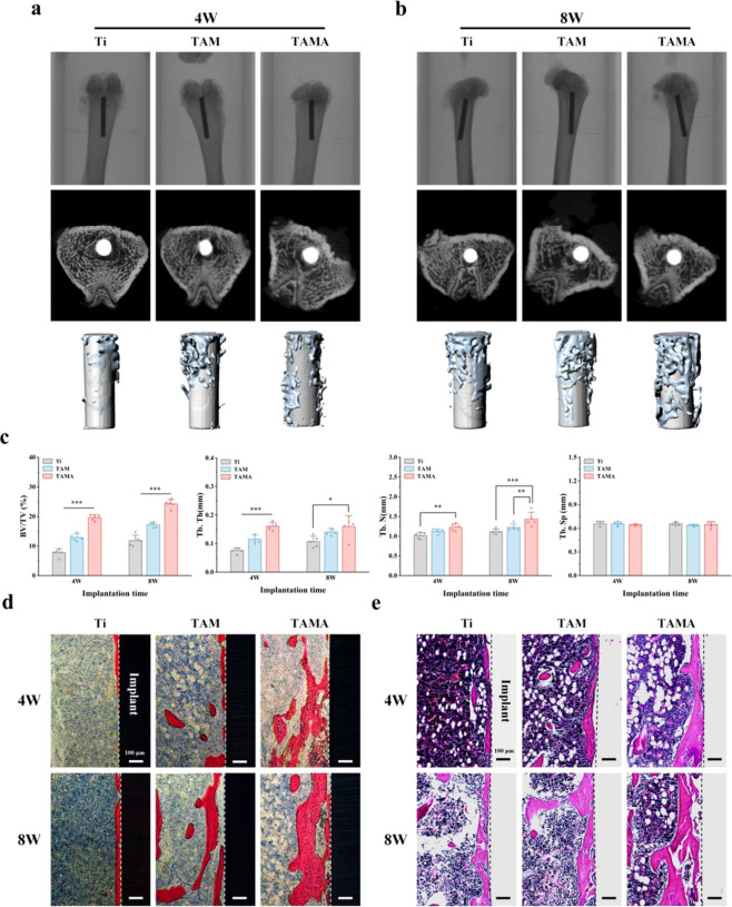
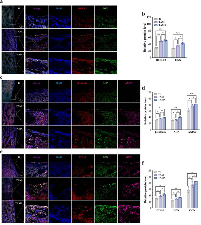
Enhanced bone healing within 1 week after post-titanium (Ti) dental implant surgery especially contributes to the subsequent long-term osseointegration, and the commonly used drug-loaded TiO2 nanotubes (TNTs) can promote osteogenesis yet still face the challenge of burst drug release that makes it difficult to maintain long-term effective drug concentrations and good osseointegration. Here, we prepared a double drug loading/release system of silk fibroin/mesoporous silica nanoparticles (SF/MSN) nanocomposite coating modified TNTs (TAMA) with AZD2858 (Wnt/β-catenin pathway agonist for promoting osteogenesis) as the therapeutic drug, realizing a long-term stable drug release and better osteogenesis. The increased β-sheet content of SF reduced the degradation rate of the SF/MSN coating, thus avoiding the AZD2858 burst release. The adsorption of MSN maintained the effective drug concentration more than 1 week that was especially critical for early bone healing. Under the protection of SF/MSN coating, the TAMA implant showed a well-organized spatial release of AZD2858, well enabling the osteogenic differentiation and mineralization at cellular level for up to 21 days. Animal experiments further demonstrated that the slow release of AZD2858 in the TAMA implant effectively activated the Wnt/β-catenin pathway, enabling rapid bone healing in the early stage of implantation and finally achieving the best osseointegration efficacy. Thus, this study proposed an efficient strategy for developing high-performance dental implants via the construction of a biodegradable SF/MSN coating.
Keywords: TiO2 nanotubes; coating; dental implant; drug delivery; osseointegration.
Figures
Similar articles
-
Silk fibroin coated TiO2 nanotubes for improved osteogenic property of Ti6Al4V bone implants.Mater Sci Eng C Mater Biol Appl. 2019 Dec;105:109982. doi: 10.1016/j.msec.2019.109982. Epub 2019 Jul 17. Mater Sci Eng C Mater Biol Appl. 2019. PMID: 31546427
-
Macroporous coating of silver-doped hydroxyapatite/silica nanocomposite on dental implants by EDTA intermediate to improve osteogenesis, antibacterial, and corrosion behavior.Biomed Mater. 2025 Jan 24;20(2). doi: 10.1088/1748-605X/ad971d. Biomed Mater. 2025. PMID: 39586175
-
Enhanced Osseointegration of Titanium Implants by Surface Modification with Silicon-doped Titania Nanotubes.Int J Nanomedicine. 2020 Nov 3;15:8583-8594. doi: 10.2147/IJN.S270311. eCollection 2020. Int J Nanomedicine. 2020. PMID: 33173295 Free PMC article.
-
Application of silk fibroin coatings for biomaterial surface modification: a silk road for biomedicine.J Zhejiang Univ Sci B. 2023 Oct 20;24(11):943-956. doi: 10.1631/jzus.B2300003. J Zhejiang Univ Sci B. 2023. PMID: 37961798 Free PMC article. Review.
-
Targeted Drug Delivery from Titanium Implants: A Review of Challenges and Approaches.Adv Exp Med Biol. 2020;1251:1-17. doi: 10.1007/5584_2019_447. Adv Exp Med Biol. 2020. PMID: 31768968 Review.
Cited by
-
Advances in Nanotechnology Research in Food Production, Nutrition, and Health.Nutrients. 2025 Jul 26;17(15):2443. doi: 10.3390/nu17152443. Nutrients. 2025. PMID: 40806027 Free PMC article. Review.
References
-
- Geng Z., Li X., Ji L., Li Z., Zhu S., Cui Z., Wang J., Cui J., Yang X., Liu C.. A novel snail-inspired bionic design of titanium with strontium-substituted hydroxyapatite coating for promoting osseointegration. J. Mater. Sci. Technol. 2021;79:35–45. doi: 10.1016/j.jmst.2020.11.041. - DOI
MeSH terms
Substances
LinkOut - more resources
Full Text Sources
Research Materials
Miscellaneous
