. 2025 Aug;73(8):1673-1691.
doi: 10.1002/glia.70026. Epub 2025 Apr 28.

Astroglial CB1 Reveal Sex-Specific Synaptic Effects of Amphetamine

Affiliations

Astroglial CB1 Reveal Sex-Specific Synaptic Effects of Amphetamine

Yamuna Mariani et al. Glia. 2025 Aug.

Abstract

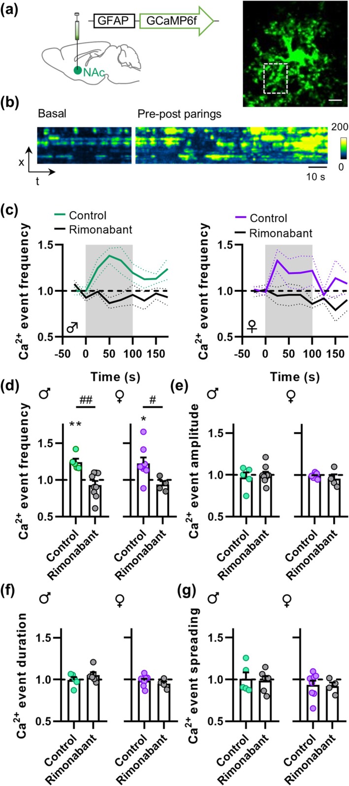
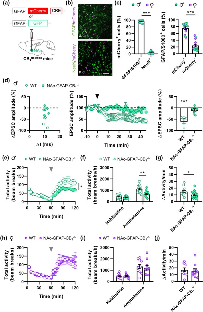
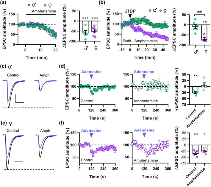
The Nucleus Accumbens (NAc) is a critical brain region for the effects of psychostimulant drugs. Type-1 cannabinoid receptors (CB1), the main elements of the endocannabinoid system (ECS) in the brain, participate in these effects and modulate synaptic functions in the NAc. Besides their neuronal expression, CB1 receptors are also present in astrocytes, where they contribute to the regulation of synaptic plasticity and behavior. However, the impact of astroglial CB1 receptors on synaptic plasticity in the NAc and on psychostimulant-induced synaptic and behavioral effects is currently unknown. This study shows that the psychostimulant amphetamine impairs a form of astroglial CB1 receptor-dependent synaptic plasticity in the NAc of male, but not female mice. Consistently, locomotor effects of amphetamine require astroglial CB1 receptors in male, but not female mice. These results, by revealing unforeseen mechanisms underlying sex-dependent effects of amphetamine, pave the way to a better understanding of the diverse impact of psychostimulants in women and men.

Keywords: CB1; adenosine; amphetamine; astrocytes; sex differences; spike‐timing‐dependent plasticity.


Conflict of interest statement

The authors declare no conflicts of interest.

Figures

FIGURE 1
FIGURE 1
Astrocytes mediate t‐LTD in the NAc. (a) Representative trace of the pre‐post pairing protocol with Δt ≈ 10 ms. (b) Change in EPSC amplitude vs. the pre‐post time windows. (c) Time course of the t‐LTD. Insets show representative traces before (black) and 45 min after STDP induction (green). Scale bars: 50 pA, 20 ms. (d) and (e) Change in EPSC amplitude after t‐LTD induction in different experimental conditions in males (d) and females (e). (f) Squared coefficient of variation (CV2) analysis. (g) and (h) Change in EPSC amplitude versus the pre‐post time windows (left), time course of t‐LTD (middle), and change in EPSC amplitude after t‐LTD induction (right) in different experimental conditions in males (g) and females (h). Arrowhead indicates t‐LTD induction. Data are expressed as mean ± s.e.m. Statistical differences comparing baseline with 45 min after t‐LTD induction (paired Student's t‐test) are expressed as (*) p < 0.05, (***) p < 0.001.
FIGURE 2
FIGURE 2
t‐LTD in the NAc requires astroglial pmCB1 receptors and adenosine release. (a) Change in EPSC amplitude vs. the pre‐post time windows (left), t‐LTD time course (middle), and change in EPSC amplitude after t‐LTD induction (right) in DN22‐CB1‐KI male mice and hemopressin. (b)–(d) Like (a) but for Nex‐CB1 −/− male mice (b) and GFAP‐CB1 −/− male (c) or female (d) mice in control conditions or when adenosine was applied. Note that Adenosine (250 μM) was puff applied through a glass pipette during t‐LTD induction. Black arrowhead, t‐LTD induction. Blue arrowhead, puff application of adenosine. Data are expressed as mean ± s.e.m. Statistical differences comparing baseline with 45 min after t‐LTD induction (paired Student's t‐test) are expressed as (*) p < 0.05, (**) p < 0.01, and (***) p < 0.001. Statistical differences comparing the different experimental groups (Two‐way ANOVA) are expressed as (##) p < 0.01, and (###) p < 0.001.
FIGURE 3
FIGURE 3
Astrocytes respond with cytosolic Ca2+ increases to t‐LTD induction. (a) Scheme depicting viral injection in the NAc (left) and maximal fluorescence projection of a representative astrocyte expressing GCaMP6f (right). Scale bar: 10 μm. (b) Fluorescence intensity over time of the astrocyte region boxed in (a) during basal conditions and during t‐LTD induction (pre‐post parings). (c) Normalized Ca2+ event frequency in control conditions and in the presence of Rimonabant. Gray area indicates t‐LTD induction. (d)–(g) Normalized Ca2+ event frequency, amplitude, duration and spreading during t‐LTD induction in control conditions and with Rimonabant. Data are expressed as mean ± s.e.m. Statistical differences comparing baseline with t‐LTD induction (paired Student's t‐test) are expressed as (*) p < 0.05, (**) p < 0.01. Statistical differences comparing Ca2+ dynamics in control conditions and Rimonabant (unpaired Student's t‐test) are expressed as (#) p < 0.05, (##) p < 0.01.
FIGURE 4
FIGURE 4
Astroglial CB1 participates in amphetamine‐induced hyperlocomotion in male mice. (a) Scheme depicting the injection of the viral vectors. (b) IHC of viral expression (mCherry) in astrocytes (GFAP) but not in neurons (NeuN). a.c.: Anterior commissure. Scale bar: 50 μm. (c) Percentage of mCherry+ cells that co‐express GFAP and/or S100β or NeuN (left) and percentage of GFAP/S100β+ cells that co‐expressed mCherry (right). (d) Change in EPSC amplitude vs. the pre‐post time windows (left), time course of t‐LTD (middle), and change in EPSC amplitude after t‐LTD induction (right). Black arrowhead indicates t‐LTD induction. (e) Time course of locomotor activity in male mice. Gray arrowhead indicates amphetamine injection. (f) Total locomotor activity during habituation and after amphetamine injection in male mice. (g) Change in activity after amphetamine injection in male mice. (h–j) Like e‐g but for female mice. Data are expressed as mean ± s.e.m. Statistical differences comparing baseline with 45 min after t‐LTD induction (paired Student's t‐test) or genotype effects on amphetamine‐induced locomotion (Two‐way ANOVA) are expressed as (*) p < 0.05, (**) p < 0.01, and (***) p < 0.001.
FIGURE 5
FIGURE 5
In vivo amphetamine treatment decreases neurotransmission and occludes t‐LTD in male but not in female mice. (a) Scheme of the experimental design. (b) t‐LTD time course and (c) change in EPSC amplitude after t‐LTD induction after amphetamine treatment in males and females. Black arrowhead indicates t‐LTD induction. (d) Representative traces (top) and sEPSC frequency and amplitude (bottom) obtained from male mice treated with saline or amphetamine. (e) Like (d) but for females. (f) and (g) Like (e) but for GFAP‐CB1 −/− male mice. Scale bars: 10 pA, 50 ms. Data are expressed as mean ± s.e.m. Statistical differences comparing baseline with 45 min after t‐LTD induction (paired Student's t‐test) and amphetamine effects in sEPSCs (unpaired Student's t‐test for panel d and e, and Two‐way ANOVA for panel g) are expressed as (*) p < 0.05, (**) p < 0.01. Statistical differences comparing t‐LTD in saline and amphetamine‐treated mice (unpaired Student's t‐test) are expressed as (#) p < 0.05.
FIGURE 6
FIGURE 6
Amphetamine occludes A1 receptors and t‐LTD in males but not in females. (a) Time course (left) and changes in EPSC amplitude (right) when amphetamine was added to the bath. (b) t‐LTD time course (left) and change in EPSC amplitude (right) after t‐LTD induction in the presence of amphetamine in males and females. Black arrowhead indicates t‐LTD induction. (c) Representative EPSC traces obtained from male mice before (black) and after adenosine application (blue) in control and in the presence of amphetamine. Scale bars: 100 pA, 20 ms. (d) Time course and change in EPSC amplitude in the different experimental conditions in male mice. (e) and (f) Like (c) and (d) but for female mice. Blue arrowheads indicate adenosine puff application. Data are expressed as mean ± s.e.m. Statistical differences comparing baseline with 45 min after t‐LTD induction and basal with adenosine application (paired Student's t‐test) are expressed as (*) p < 0.05, (**) p < 0.01 and (***) p < 0.001. Statistical differences comparing t‐LTD in males and females (unpaired Student's t‐test) are expressed as (##) p < 0.01.
FIGURE 7
FIGURE 7
Male mice show higher sensitivity to adenosine. (a) Representative EPSC traces obtained from male and female mice in response to increasing concentrations of adenosine. (b) Change in EPSC amplitude in response to increasing concentrations of adenosine. (c) Paired‐pulse ratio before and after adenosine application. (d) Representative EPSC traces obtained from male and female mice before (basal) and after CPT (2 μM) perfusion. (e) Time course and (f) change in EPSC amplitude in response to CPT in males and females. Data are expressed as mean ± s.e.m. Statistical differences comparing adenosine dose–response (Two‐way ANOVA) and CPT (unpaired Student's t‐test) effects in males and females and paired‐pulse ratio before and after adenosine (paired Student's t‐test) are expressed as (**) p < 0.01, and (***) p < 0.001. Scale bars: 100 pA, 20 ms.

References

    1. Ahn, K. C. , Bernier B. E., Harnett M. T., and Morikawa H.. 2010. “IP3 Receptor Sensitization During In Vivo Amphetamine Experience Enhances NMDA Receptor Plasticity in Dopamine Neurons of the Ventral Tegmental Area.” Journal of Neuroscience 30, no. 19: 6689–6699. 10.1523/JNEUROSCI.4453-09.2010. - DOI - PMC - PubMed
    1. Andrade‐Talavera, Y. , Duque‐Feria P., Paulsen O., and Rodriguez‐Moreno A.. 2016. “Presynaptic Spike Timing‐Dependent Long‐Term Depression in the Mouse Hippocampus.” Cerebral Cortex 26, no. 8: 3637–3654. 10.1093/cercor/bhw172. - DOI - PMC - PubMed
    1. Araque, A. , Carmignoto G., Haydon P. G., Oliet S. H., Robitaille R., and Volterra A.. 2014. “Gliotransmitters Travel in Time and Space.” Neuron 81, no. 4: 728–739. 10.1016/j.neuron.2014.02.007. - DOI - PMC - PubMed
    1. Araque, A. , Castillo P. E., Manzoni O. J., and Tonini R.. 2017. “Synaptic Functions of Endocannabinoid Signaling in Health and Disease.” Neuropharmacology 124: 13–24. 10.1016/j.neuropharm.2017.06.017. - DOI - PMC - PubMed
    1. Araque, A. , Parpura V., Sanzgiri R. P., and Haydon P. G.. 1999. “Tripartite Synapses: Glia, the Unacknowledged Partner.” Trends in Neurosciences 22, no. 5: 208–215. 10.1016/s0166-2236(98)01349-6. - DOI - PubMed

Substances

LinkOut - more resources