

Long-Term Impact of Intralesional Bony Overgrowth on Opposing Cartilage Integrity: Five-Year Results Following Cartilage Repair
- PMID: 40289954
- PMCID: PMC12037522
- DOI: 10.1177/19476035251335008
Long-Term Impact of Intralesional Bony Overgrowth on Opposing Cartilage Integrity: Five-Year Results Following Cartilage Repair
Abstract
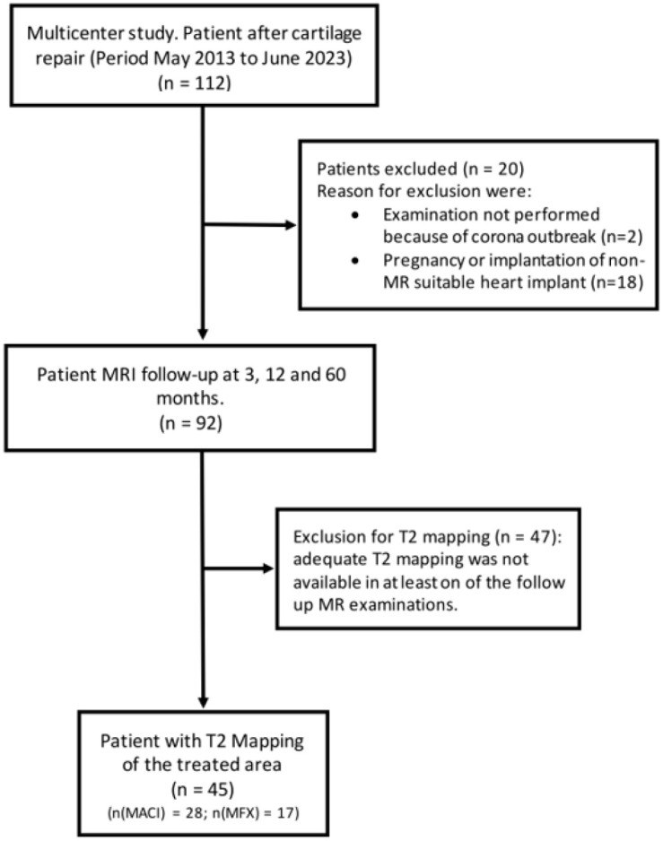
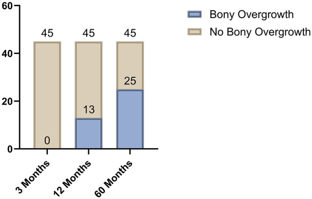
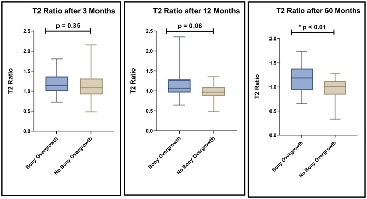
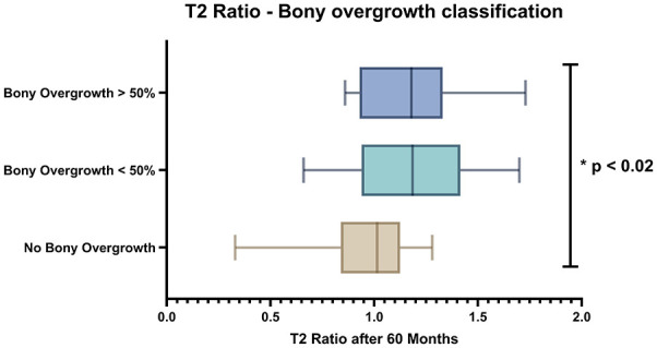
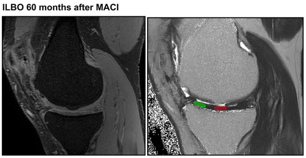
ObjectivesThis study aimed to assess the impact of intralesional bony overgrowth (ILBO) after cartilage repair on the integrity of opposing articulating cartilage (OpAC) using T2 mapping and to correlate these findings with clinical outcomes.MethodsIn this multicenter study, magnetic resonance imaging (MRI) examinations were performed in the follow-up after cartilage repair (Microfracturing (MFX) and Matrix-Induced Autologous Chondrocyte Implantation (MACI)) in 45 patients up to 5 years after surgery. T2 values of the OpAC after 3, 12, and 60 months in patients with and without ILBO after 60 months were conducted along with clinical assessments (International Knee Documentation Committee (IKDC) and Knee injury and Osteoarthritis Outcome Score (KOOS)).ResultsAt 60 months post-surgery, 44.4% of patients presented with ILBO, which was associated with significantly higher T2 values in OpAC (P = 0.004). A tendency toward increased T2 values was observed after 12 months, although this did not reach statistical significance (P = 0.06). However, no significant differences were found in clinical outcomes between patients with or without ILBO, nor between those with or without T2 values comparable to reference cartilage.ConclusionILBO significantly affects the biophysical MRI properties of OpAC as indicated by higher T2 values after 60 months. These alterations, though not reflected in any clinical score, can suggest potential long-term implications for cartilage degeneration and may inform future monitoring strategies for cartilage repair. Further research is required to evaluate the long-term effects of these altered mechanical impacts on articulating cartilage and their clinical implications.
Keywords: articular cartilage; bone formation; follow-up studies; knee joint; magnetic resonance imaging; subchondral arthroplasty.
Conflict of interest statement
The author(s) declared no potential conflicts of interest with respect to the research, authorship, and/or publication of this article.
Figures

Similar articles
-
Long-term Assessment of Subchondral Bone Changes and Intralesional Bony Overgrowth After Third-Generation Autologous Chondrocyte Implantation: A Retrospective Study.Am J Sports Med. 2023 May;51(6):1414-1421. doi: 10.1177/03635465231162107. Epub 2023 Apr 18. Am J Sports Med. 2023. PMID: 37070725 Free PMC article.
-
Bone marrow edema-like signal after cartilage repair does not affect outcomes in a five-year follow-up.Eur Radiol. 2025 Apr;35(4):1808-1817. doi: 10.1007/s00330-024-11078-8. Epub 2024 Sep 16. Eur Radiol. 2025. PMID: 39285030 Free PMC article.
-
Cartilage T2 Relaxation Times and Subchondral Trabecular Bone Parameters Predict Morphological Outcome After Matrix-Associated Autologous Chondrocyte Implantation With Autologous Bone Grafting.Am J Sports Med. 2020 Dec;48(14):3573-3585. doi: 10.1177/0363546520965987. Epub 2020 Nov 17. Am J Sports Med. 2020. PMID: 33200942
-
Marrow Stimulation Has Relatively Inferior Patient-Reported Outcomes in Cartilage Restoration Surgery of the Knee: A Systematic Review and Meta-analysis of Randomized Controlled Trials.Am J Sports Med. 2022 Mar;50(3):858-866. doi: 10.1177/03635465211003595. Epub 2021 Apr 23. Am J Sports Med. 2022. PMID: 33890799
-
Comparative Effectiveness of Cartilage Repair With Respect to the Minimal Clinically Important Difference.Am J Sports Med. 2019 Nov;47(13):3284-3293. doi: 10.1177/0363546518824552. Epub 2019 May 13. Am J Sports Med. 2019. PMID: 31082325
References
LinkOut - more resources
Full Text Sources
