

Metastatic pheochromocytoma complicated with Langerhans cell histiocytosis: a case report
- PMID: 40290304
- PMCID: PMC12021616
- DOI: 10.3389/fendo.2025.1494783
Metastatic pheochromocytoma complicated with Langerhans cell histiocytosis: a case report
Abstract
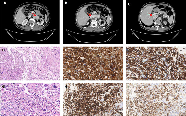
Pheochromocytoma is a neuroendocrine neoplasm that originates from chromaffin cells of the adrenal medulla. Langerhans cell histiocytosis (LCH) is a proliferative disease of histiocyte-like cells, often associated with activating mutations of the mitogen-activated protein kinase (MAPK) pathway. We present a case of a 49-year-old male with a history of pheochromocytoma, which metastasized to the inferior vena cava eight years after left adrenalectomy. At the same time, it was found that the pheochromocytoma in the metastasis was complicated with LCH, a combination that has not been previously reported. Genetic analysis was carried out by next-generation sequencing (NGS) technology. Somatic mutations of BRAF and RAD54B were detected in Langerhans cells and EPAS1 in pheochromocytoma.
Keywords: EPAS1 gene; Langerhans cell histiocytosis; case report; metastasis; pheochromocytoma.
Copyright © 2025 Dai and Xie.
Conflict of interest statement
The authors declare that the research was conducted in the absence of any commercial or financial relationships that could be construed as a potential conflict of interest.
Figures

Similar articles
-
A giant cystic pheochromocytoma of the adrenal gland.Endocr Pathol. 2008 Summer;19(2):133-8. doi: 10.1007/s12022-008-9016-4. Endocr Pathol. 2008. PMID: 18322657
-
Coexistence of neuroblastoma detected on staging of Langerhans cell histiocytosis.Pediatr Int. 2014 Aug;56(4):608-10. doi: 10.1111/ped.12292. Pediatr Int. 2014. PMID: 25252048
-
Surgical management of pheochromocytoma in children.J Pediatr Surg. 1974 Apr;9(2):179-84. doi: 10.1016/s0022-3468(74)80118-1. J Pediatr Surg. 1974. PMID: 4825789 No abstract available.
-
Preoperative and surgical therapy in sporadic and familial pheochromocytoma.Front Horm Res. 2004;31:121-44. doi: 10.1159/000074661. Front Horm Res. 2004. PMID: 14674308 Review. No abstract available.
-
Cutaneous adult xanthogranuloma with a small portion of BRAFV 600E mutated Langerhans cell histiocytosis populations: A case report and the review of published work.J Dermatol. 2019 Feb;46(2):161-165. doi: 10.1111/1346-8138.14725. Epub 2018 Dec 7. J Dermatol. 2019. PMID: 30536719 Review.
References
Publication types
MeSH terms
LinkOut - more resources
Full Text Sources
Medical
Research Materials