Review
. 2025 Apr 11:16:1553794.
doi: 10.3389/fendo.2025.1553794. eCollection 2025.

Molecular mechanisms and targeted therapy of progranulin in metabolic diseases

Affiliations
Review

Molecular mechanisms and targeted therapy of progranulin in metabolic diseases

Xiaxia Wang et al. Front Endocrinol (Lausanne). .

Abstract

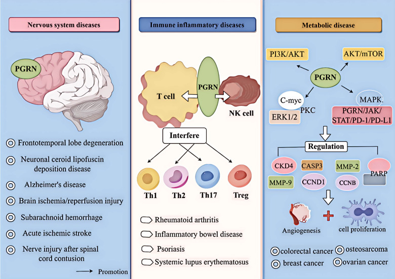
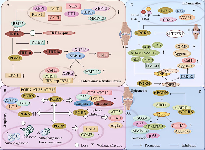
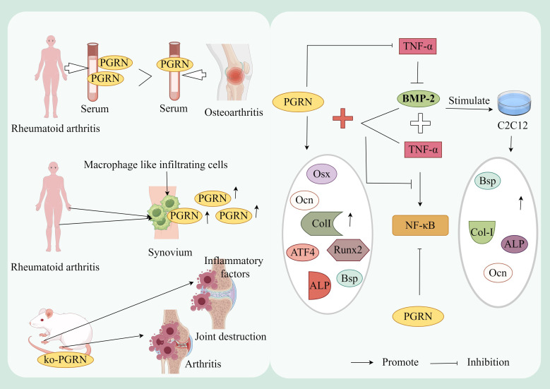
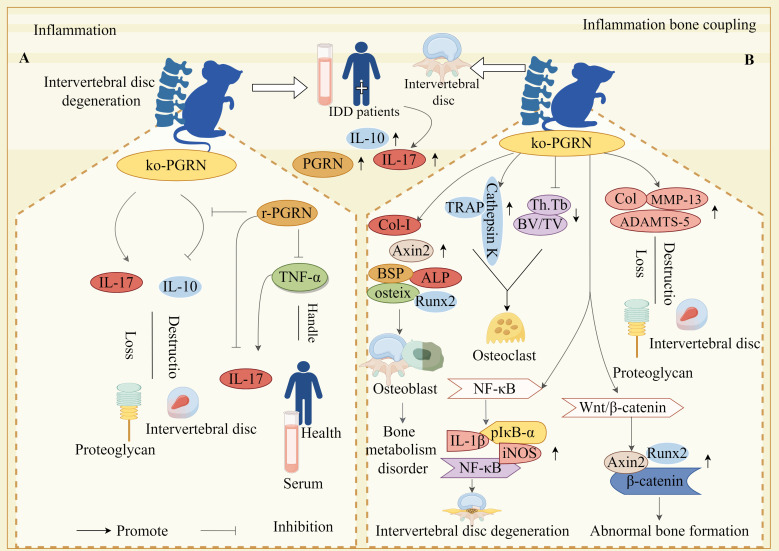
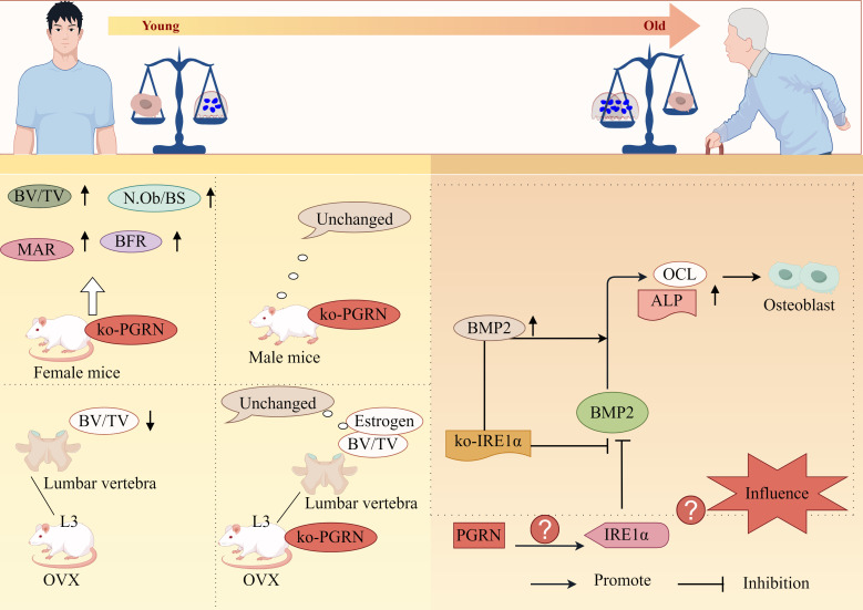
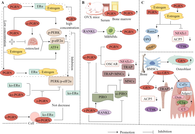
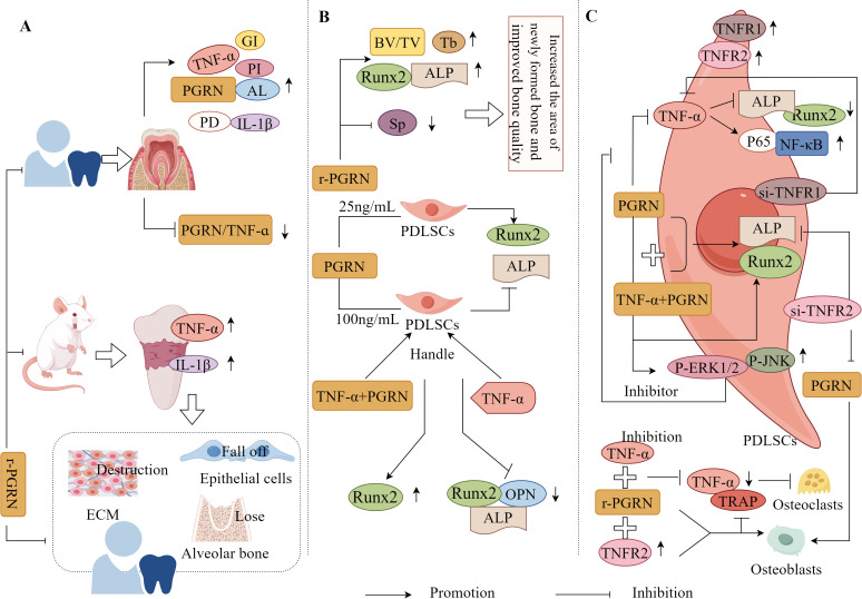
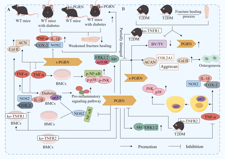
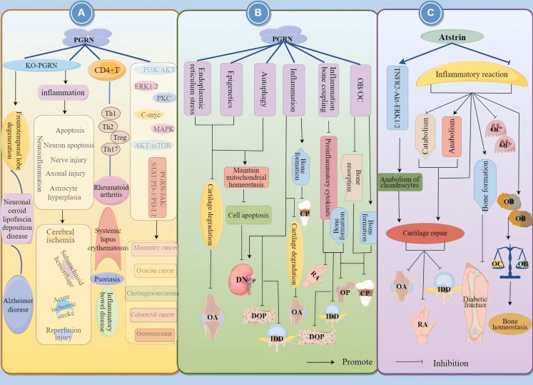
Progranulin (PGRN) is a secreted glycoprotein with cytokine-like properties, exerting tripartite mechanisms of inflammation suppression, tissue repair promotion, and metabolic regulation. This multifaceted functionality positions PGRN as a potential "multi-effect therapeutic strategy" for metabolic disorders characterised by cartilage degradation and imbalanced bone remodelling, potentially establishing it as a novel therapeutic target for such conditions. Osteoarthritis, rheumatoid arthritis, intervertebral disc degeneration, osteoporosis, periodontitis, and diabetes-related complications-representing the most prevalent metabolic diseases-currently lack effective treatments due to incomplete understanding of their precise pathogenic mechanisms. Recent studies have revealed that PGRN expression levels are closely associated with the onset and progression of these metabolic disorders. However, the exact regulatory role of PGRN in these diseases remains elusive, partly owing to its tissue-specific actions and context-dependent dual roles (anti-inflammatory vs. pro-inflammatory). In this review, we summarise the structure and functions of PGRN, explore its involvement in neurological disorders, immune-inflammatory diseases, and metabolic conditions, and specifically focus on its molecular mechanisms in metabolic diseases. Furthermore, we consolidate advances in targeting PGRN and the application of its engineered derivative, Atsttrin, in metabolic bone disorders. We also discuss potential unexplored mechanisms through which PGRN may exert influence within this field or other therapeutic domains. Collectively, this work aims to provide a new framework for elucidating PGRN's role in disease pathogenesis and advancing strategies for the prevention and treatment of metabolic disorders.

Keywords: PGRN; bone homeostasis; cartilage repair; inflammation; metabolic diseases; targeted therapy.


Conflict of interest statement

The authors declare that the research was conducted in the absence of any commercial or financial relationships that could be construed as a potential conflict of interest.

Figures

Figure 1
Figure 1
Relationship between PGRN and disease. PGRN is closely related to frontotemporal lobe degeneration, neuronal ceroid lipofuscin deposition disease, Alzheimer’s disease, Brain ischemia/reperfusion injury, subarachnoid haemorrhage, acute ischemic stroke and nerve injury after spinal cord contusion; PGRN is associated with rheumatoid arthritis, inflammatory bowel disease, psoriasis and systemic lupus erythematosus by interfering with T cells (affecting the proliferation and differentiation of Th1, Th2, Th17, Treg, etc.) and NK cells. PGRN mediates PI3K/Akt ERK1/2, PKC, C-myc, MAPK, AKT/mTOR, PGRN/JAK/STAT/PD-1/PD-L1, it can regulate the activity of CKD4 and the levels of CCNB and CCND1, up regulate MMP-9 and activate MMP-2, regulate the cleavage of CASP3 and PARP, and induce the angiogenesis, proliferation, invasion and migration of cancer cells, which is closely related to the development of cancer diseases such as colorectal cancer, osteosarcoma, breast cancer and ovarian cancer.
Figure 2
Figure 2
Molecular mechanism of PGRN in osteoarthritis. (A) IRE1α overexpression or BMP2 treatment reduced the expression of Col II, Sox9, Col X, MMP-13, Runx2, IHH and increased the expression of PTHrP, while IRE1 α point mutant (IRE1α-PM) completely lost these regulatory activities of IRE1α. PGRN combines with the kinase domain of IRE1α during endoplasmic reticulum stress, assists in IRE1α phosphorylation, promotes XBP1u splicing to generate XBP1s, causes IRE1α to undergo nuclear translocation, and up regulates the expression of Col II. In addition, ERN1 was positively correlated with the level of PGRN. ERN1 deficiency reduced the protein levels of Col II, PGRN, XBP1s and IRE1α/p-IRE1α and increased the number of MMP-13 positive cells by reducing the expression of PGRN and XBP1 splicing, resulting in abnormal collagen structure, which accelerated cartilage degeneration and homeostasis imbalance in OA. (B) PGRN deletion significantly increased the expression levels of autophagy markers LC3-II, P62 and ATG5-ATG12 complex, but PGRN deletion did not affect the mRNA levels of LC3, p62 and ATG12. PGRN overexpression increased the level of autophagosome markers and lysosome number, but did not promote autophagosome formation and lysosome biogenesis. PGRN treatment reduced the expression of Col X and COMP, while PGRN and autophagy inhibitor CO treatment down regulated the autophagy marker ATG5, Expression of LC3II and ATG12. PGRN deficiency reduced the interaction between PGRN and PGRN-ATG5-ATG12, increased the expression of P62, Caspase-3 and Caspase-12 in chondrocytes, and decreased the proliferation, increased apoptosis and impaired autophagy activation of chondrocytes. (C) The expression of PGRN mRNA and the expression levels of no, COX-2, MMP13 and VCAM-1 in differentiated chondrocytes treated with TNF-α, IL-1β, IL-6 and TLR-4 agonists were significantly increased, while rpgrn significantly inhibited the results induced by inflammatory factors. Under the stimulation of IL-1β or LPS, si-TNFR1 inhibited the anti-inflammatory effect of PGRN. In PGRN deficient mice, the cartilage degeneration related factors ALP, BGP, MMP-13, ADAMTS-5, ADAMTS-7, ADAMTS-12, iNOS and COX-2 were significantly increased, and the degradation rate of COMP and Aggrecan was accelerated, which was reversed by rPGRN. In addition, in a TNFR2 dependent manner, PGRN, on the one hand, increased the levels of anabolic biomarkers Col II and Aggrecan by activating ERK1/2 signalling pathway, and restored the metabolic balance of chondrocytes. On the other hand, PGRN inhibited the expression levels of MMP13, ADAMTS-5, COX-2 and iNOS and the excessive degradation of Col II and Aggrecan by interacting with TNF-α, blocking the progress of OA. (D) PGRN enhanced the expression and activity of SIRT1 in a dose-dependent manner, prevented P65 nuclear translocation through p65 deacetylation, inhibited TNF-α induced p65 nuclear accumulation, significantly increased the expression and secretion of Col IIA1 and Aggrecan, and decreased the expression of total p65 and acetylated P65, MMP-13, and ADAMTS-5 in chondrocytes induced by TNF-α. The effect of PGRN was reversed by the CO treatment of rPGRN and si-SIRT1.
Figure 3
Figure 3
Mechanism of PGRN in rheumatoid arthritis. The levels of PGRN in serum and synovial fluid of RA patients were significantly higher than those of OA patients, and the synovial macrophage like infiltrating cells of RA patients strongly expressed PGRN. In addition, PGRN deficient mice immunised with type II collagen developed more severe inflammatory arthritis and bone and joint damage. The expression of OB differentiation marker genes CoL-I, OCN and BSP and ALP activity were significantly increased after BMP-2 stimulated mouse myoblasts (C2C12). TNF-α treatment reversed this effect of BMP-2. PGRN treatment blocked TNF-α mediated inhibition of ALP activity and mineralisation. Compared with the cells treated with TNF-α and BMP-2 alone, the mRNA expression of OB specific marker genes Col I, OCN, BSP, Runx2, OSX and ATF4, ALP activity and mineralisation of cells treated with PGRN were significantly increased. In addition, the activity of NF-κB was significantly increased after TNF-α+BMP-2 treatment, while the expression of NF-κB was inhibited in a concentration dependent manner after TNF-α+BMP-2+PGRN treatment. In addition, the activity of NF-κB was significantly decreased in cells treated with PGRN alone.
Figure 4
Figure 4
Molecular mechanism of PGRN in intervertebral disc degeneration. (A) The expressions of PGRN, IL-10 and IL-17 in peripheral blood and intervertebral disc tissue of IDD patients were significantly increased. In the animal model, the loss of PGRN led to the decrease of IL-10 expression and the increase of IL-17 expression, which accelerated the loss of proteoglycan and the destruction of intervertebral disc. After stimulation with r-pgrn, the transcription level of IL-10 increased significantly. TNF-α significantly increased the transcription and protein levels of IL-17, while r-pgrn significantly inhibited the expression of IL-17 induced by TNF-α. (B) The expression levels of OB marker genes ALP, osteix, CoL-I, BSP, AXIN2 and Runx2 were significantly increased in PGRN knockout mice, Bone formation in IVD. The levels of Col, MMP-13 and ADAMTS-5 in cartilage tissue of PGRN-deficient mice were significantly increased, and proteoglycan loss and cartilage degradation in endplate cartilage were serious. The expressions of TRAP and Cathepsin K were significantly increased in the new osteoblasts, BV/TV and Th.Tb decreased significantly, The activity of OC increased. In addition, on the one hand, PGRN deficiency caused the over activation of NF-κB signalling pathway, and the levels of NF-κB, PIκB-α, IL-1β and iNOS were significantly increased, which led to the accelerated degradation of IVD. On the other hand, PGRN deficiency caused the activation of Wnt/β-catenin signalling pathway, and the levels of AXIN2, Runx2 and β-Catenin were significantly increased, which induced abnormal bone formation.
Figure 5
Figure 5
PGRN regulates osteoporosis progression by regulating OB differentiation. Bv/TV, N.Ob/BS, MAR and BFR were significantly increased in PGRN knockout female mice, but not in male mice. The BV/TV of the third lumbar vertebra (L3) cancellous bone in OVX WT mice was significantly decreased, while the BV/TV of the third lumbar vertebra (L3) cancellous bone and oestrogen depletion in OVX ko-PGRN mice were not significantly changed. BMP2 leads to high ALP activity and strong OCL production, and promotes osteogenic differentiation. IRE1α overexpression inhibits BMP2 mediated osteogenic differentiation. IRE1α knockdown can significantly increase the expression of BMP2. ko-IRE1α can increase the expression of ALP and OCL induced by BMP2, and stimulate the differentiation of OB induced by BMP2. Whether PGRN can interfere with OB differentiation and proliferation by regulating the expression of IRE1α has not been reported.
Figure 6
Figure 6
Molecular mechanism of PGRN in osteoporosis. (A) PGRN and E2 could significantly inhibit OC activity, while si-PGRN could significantly induce OC activity. At low E2 concentrations, PGRN acts as a chaperone protein to assist the binding of E2 to ERα. However, at high E2 concentrations, PGRN cannot play a normal molecular chaperone role, interfering with the binding of E2 to ERα. The effects of PGRN, E2 and PGRN+E2 on promoting osteogenesis and inhibiting OC were significantly weakened after ko-ERα. PGRN, E2 and PGRN+E2 could significantly up regulate the levels of p-PERK, p-eIF2α and ATF4, and activate the PERK/p-eIF2α signalling pathway. After ko-ERα and ko-PGRN, the results were opposite, while r-PGRN could partially restore the function of PERK/p-eIF2α signalling pathway. In addition, in ko-PGRN cells, after ko-ERα, r-PGRN treatment did not significantly reduce PERK/p-eIF2α signal transduction. (B) The levels of PGRN in serum and bone marrow fluid of OVX mice were significantly increased. In RANKL treated mbmms, PGRN mRNA and protein levels were significantly increased in a time-dependent manner. si-PGRN significantly inhibited the gene expression of PGRN, down regulated the expression levels of TRAP, NFATc1 and OSCAR, and reduced the number of TRAP+MNCs. In the presence of RANKL, r-PGRN reversed the above results, and PGRN significantly increased the expression of PIRO, ko-PGRN could significantly reduce the expression of PIRO, while si-PIRO significantly inhibited the formation of MNCs, reduced the number of TRAP+MNCs and PIRO mRNA level, r-PGRN partially restored MNCs inhibited by PIRO transfection. (C) PGRN could significantly increase the level of Estrogens, and PGRN and Estrogens could up regulate the expression of osteogenic markers Runx2, OPG and DMP1, and down regulate the expression of OC related markers NFATc1, ACP5 and CTSK. When PGRN+Estrogens co acted, the regulatory effect was more obvious. GRN was expressed in mouse bone tissue and cultured OB from BMSCs. ko-GRN led to a significant reduction in GRN mRNA levels, OC markers CTSK and ACP5 expression in bone and cultured OB from male and female mice with ko-PGRN. In contrast, the expression of TRAP-positive cells, immature OC-associated genes Csf1r, Adgre1 and C1q, and the expression of osteogenesis-related factors C3 and Osm in mouse OC were increased in distal femoral trabeculae.
Figure 7
Figure 7
Molecular mechanism of PGRN in periodontitis. (A) In CP patients, PI, PD, Al, GI, PGRN, TNF-α and IL-1β in gingiva and gingival crevicular fluid were significantly increased, and PGRN/TNF-α was significantly decreased. The expression of TNF-α and IL-1β in the connective tissue of EP rats were significantly increased, accompanied by ECM destruction, partial epithelial cell loss, and significant loss of alveolar bone. Local treatment with r-PGRN inhibited the inflammatory reaction of periodontal tissue and alleviated the progress of periodontitis. (B) r-PGRN can significantly increase the BV/TV and TB values and the expression of ALP and Runx2 in periodontitis rats, reduce the SP value, increase the area of new bone in periodontitis rats, and improve the bone quality. In addition, 25 ng/ml PGRN promoted the proliferation of PDLSCs, significantly increased the expression of ALP and Runx2 mRNA, and promoted the differentiation of OB, while 100 ng/ml PGRN significantly decreased the expression of ALP and Runx2 mRNA. The mRNA and protein expressions of ALP, Runx2 and OPN in PDLSCs treated with TNF-α were significantly decreased, while the mRNA and protein levels of Runx2 in hPDLSCs treated with TNF-α+PGRN were significantly increased. (C) TNFR1 and TNFR2 were positive in hPDLSCs. TNF-α stimulation could significantly reduce the expression of ALP and Runx2 in hPDLSCs, and PGRN treatment could significantly reverse the effect of TNF-α. In hPDLSC cells si-TNFR1, the inhibition of osteogenesis mediated by TNF-α disappeared, but TNF-α+PGRN and PGRN alone significantly promoted the mRNA and protein expression of ALP and Runx2. In hPDLSCs cells si-TNFR2, TNF-α-mediated osteogenic inhibition was lost, but both TNF-α+PGRN and PGRN alone significantly promoted mRNA and protein expression of ALP and Runx2. PGRN significantly enhanced the expression of p-ERK1/2, p-JNK, ALP and Runx2 in hPDLSCs si-TNFR1, while ERK1/2 and JNK inhibitors significantly eliminated the expression of ALP and Runx2 enhanced by PGRN. The expression of p65 was significantly increased when hPDLSCs were stimulated with TNF-α. In hPDLSCs transfected with TNFR2, TNF-α activated NF-κB, p65 and inhibited osteogenic differentiation, which was saved by PGRN. In addition, r-PGRN and anti TNF-α treatment can significantly reduce the expression of TNF-α and the number of trap positive cells during periodontal bone defect regeneration, and inhibit OC formation. TNFR2+r-PGRN promotes OB differentiation and inhibits OC differentiation.
Figure 8
Figure 8
Molecular mechanism of PGRN in DOP. (A) Compared with non-diabetic WT mice, the fracture healing of diabetic WT mice, non-diabetic ko-PGRN mice and diabetic ko-PGRN mice was weakened, and TNF-α, IL-1β, NOS2 and COX-2 were significantly increased, especially in diabetic ko-PGRN mice. After r-PGRN treatment, the levels of IL-1β, NOS2, COX-2 and NOS2 were significantly decreased, while Col II and ACN were significantly increased. In normal BMCs, TNF-α induced phosphorylation of P38, JNK and NF-κB, which was inhibited by r-PGRN. In addition, compared with normal BMCs, pro-inflammatory signalling pathway and P65 were over activated in diabetic BMCs, and this activation was further enhanced in the presence of TNF-α, while r-PGRN inhibited TNF-α induced signal transduction in normal and diabetic cells. In normal and diabetic BMCs, PGRN effectively inhibited the activation of NF-κB signal, and the expression of NOS2 increased in the presence of TNF-α, while the up regulation decreased after r-PGRN. In diabetic BMCs, PGRN effectively inhibited the transcription levels of IL-1β, COX-2 and NOS2 induced by TNF-α. In addition, in TNFR1 deficient BMCs, PGRN significantly induced Col II and ACAN transcription levels, while in TNFR2 deficient BMCs, these effects mediated by PGRN were almost eliminated, and Akt, ERK1/2 and mTOR signals were activated by PGRN during PGRN stimulated cartilage formation. (B) In the process of fracture healing in T2DM and T2DM, PGRN was significantly increased and BV/TV was significantly decreased. After local administration of r-PGRN, BV/TV ratio was increased, COL2A1, ACAN, Col II and Aggrecan were significantly increased, and bone formation was improved. When TNF-α was present, the expressions of IL-1β, COX-2 and NOS2 were significantly increased, P65 was transferred from cytoplasm to nucleus, and JNK and P38 were activated. PGRN inhibited this reaction mediated by TNF-α, significantly increased the levels of COL2A1 and ACAN in T2DM mice, and activated Akt and ERK1/2 signalling pathways. When ko-TNFR1 was blocked, the anabolic effect of PGRN was abolished, and the effect of PGRN on the activation of Akt and ERK1/2 disappeared.
Figure 9
Figure 9
Molecular mechanisms of PGRN in diabetic nephropathy. BMI, EGFR, UACR, TNFR1 and PGRN were correlated with the occurrence of DN, and serum PGRN level was correlated with BMI, CRP, EGFR and UACR. Serum PGRN level was positively correlated with BMI, negatively correlated with EGFR, and slightly correlated with TNFR1. The level of PGRN in T2DM patients with microangiopathy gradually increased, and the values of HbA1c, FPG, FINS, HOMA-IR, TNF-α and IL-6 significantly increased, and dyslipidaemia, hypertension and central obesity occurred. In addition, serum PGRN can stimulate adipocytes to release more IL-6, while ko-PGRN can completely block IL-6 secreted by TNF-α. The level of PGRN in renal biopsy tissue of DN patients decreased. Decrease of PGRN level in kidney of HG and STZ induced diabetic mice, compared with WT type diabetic mice, PGRN deficient diabetic mice had glomerular mesangial dilation, increased cells, increased urinary protein excretion, increased capillaries and GBM foot process loss, and further decreased glomerular capillary loop slit membrane proteins Podocin and Nephrin. In diabetic mice with PGRN deficiency, caspase-3 was increased, coxiv was decreased, and mitochondrial damage was aggravated. Under HG condition, the percentage of mitochondrial fragmentation in podocytes was higher, PGC-1α was significantly reduced, r-PGRN significantly inhibited l HG induced mitochondrial division, reduced the decline rate of MMP, mtDNA: nDNA ratio, and enhanced the protein expression levels of PGC-1α and TFAM. PINK1 and PARK2 were significantly decreased under HG conditions, which was rescued by r-PGRN, and mitochondria and autophagosomes co-localised in r-PGRN-treated podocytes. In HG-treated podocytes, PGRN specifically induced the expression of SIRT1 and reduced the acetylation levels of PGC-1α and FoxO1, whereas the inhibition of SIRT1 expression counteracted the protective effect of PGRN on podocytes.
Figure 10
Figure 10
Molecular mechanisms of PGRN in diabetic nephropathy. PGRN expression was significantly reduced in the kidneys of STZ-induced diabetic mice, and PGRN deficiency resulted in significantly higher kidney weights, kidney weight to body weight ratios, dilation of glomerular tunica and capillary atrophy, thickening of the glomerular GBM, and widening and loss of podocyte pedicle synapses, which were reversed by r-PGRN. Under HG conditions, PGRN expression was reduced in human podocytes (HPC), mouse podocytes (MPC), and human renal proximal tubule HK2 cells, and cleavage of caspase-3 and PARP1 was reduced, with the opposite result for r-PGRN treatment. In vivo, ko-PGRN decreased the ratio of LC3II/I and aggravated autophagy damage in the kidneys of diabetic mice foot cells; in vitro, r-PGRN treatment was the opposite. PGRN activates AMPK signalling and inhibits mTORC1 activity in HG-treated podocytes, resulting in enhanced ULK1 Ser555 phosphorylation sites and reduced ULK1 Ser757 phosphorylation sites in podocytes, whereas inhibition of the AMPK signalling pathway impairs PGRN-rescued autophagy and podocyte survival under hyperglycaemic conditions. CAMKK is activated by PGRN in podocytes under HG condition, silencing of the CAMKK-β gene or specific inhibition of CAMKK activity reduces PGRN-mediated phosphorylation of the AMPKα Thr172 phosphorylation site and ULK1 Ser555 phosphorylation site, and BAPTA-AM chelating of the cytoplasmic calcium-free antagonises the PGRN-activated AMPKα signalling pathway.
Figure 11
Figure 11
The role and mechanisms of action of PGRN (Progranulin) and its derivative Atsttrin in disease pathogenesis. (A) KO-PGRN not only directly contributes to neurodegenerative disorders such as frontotemporal lobar degeneration, neuronal ceroid lipofuscinosis, and Alzheimer’s disease, but also induces neuroinflammation, cellular apoptosis, axonal damage, astroglial hyperplasia, and neuronal death through inflammatory responses. This cascade mechanism predisposes individuals to cerebral ischaemia/reperfusion injury, subarachnoid haemorrhage, and acute ischaemic stroke. Furthermore, PGRN plays a significant role in immune-inflammatory pathologies including rheumatoid arthritis, inflammatory bowel disease, psoriasis, and systemic lupus erythematosus by modulating the differentiation of CD4+ T cells into Th1, Th2, Th17, and Treg subsets. Additionally, PGRN mediates multiple signalling pathways - including PI3K/AKT, ERK1/2, PKC, C-myc, MAPK, AKT/mTOR, and PGRN/JAK/STAT/PD-1/PD-L1 - thereby influencing the progression of various malignancies such as colorectal carcinoma, osteosarcoma, ovarian cancer, cholangiocarcinoma, and breast cancer. (B) Furthermore, PGRN exerts protective effects against OA by suppressing cartilage degradation through endoplasmic reticulum stress, epigenetic regulation, and autophagy. PGRN also maintains mitochondrial homeostasis and inhibits cellular apoptosis via autophagy, thereby demonstrating protective functions in DN. Through inflammatory responses, PGRN exhibits dual regulatory roles: while directly promoting DN progression and suppressing IDD and DOP, it concurrently protects against OA and CP by inhibiting cartilage degradation and enhancing bone formation. Additionally, PGRN modulates the inflammation-bone coupling axis to suppress inflammatory reactions and promote osteogenesis, consequently inhibiting the development of RA, DOP, OP, and IDD. By regulating OB-OC equilibrium, PGRN inhibits bone resorption while stimulating bone formation, thereby conferring protection against CP and OP. (C) Finally, the PGRN derivative Atsttrin demonstrates therapeutic potential through dual mechanisms: it promotes anabolic processes and cartilage repair in OA, RA, and IDD by activating the TNFR2-Akt-ERK1/2 pathway while suppressing inflammatory responses and catabolic activity. Concurrently, Atsttrin enhances bone formation, inhibits OC differentiation, and modulates OB-OC balance, offering protective effects against bone destructive disorders and pathologies involving bone homeostasis dysregulation.

Similar articles

References

    1. Liao S, Zheng Q, Shen H, Yang G, Xu Y, Zhang X, et al. . HECTD1-mediated ubiquitination and degradation of rubicon regulates autophagy and osteoarthritis pathogenesis. Arthritis Rheumatol. (2023) 75:387–400. doi: 10.1002/art.42369 - DOI - PubMed
    1. Lu C, Zhao H, Liu Y, Yang Z, Yao H, Liu T, et al. . Novel role of the SIRT1 in endocrine and metabolic diseases. Int J Biol Sci. (2023) 19:484–501. doi: 10.7150/ijbs.78654 - DOI - PMC - PubMed
    1. Marafie SK, Al-Mulla F, Abubaker J. mTOR: its critical role in metabolic diseases, cancer, and the aging process. Int J Mol Sci. (2024) 25:6141. doi: 10.3390/ijms25116141 - DOI - PMC - PubMed
    1. Valdez C, Wong YC, Schwake M, Bu G, Wszolek ZK, Krainc D. Progranulin-mediated deficiency of cathepsin D results in FTD and NCL-like phenotypes in neurons derived from FTD patients. Hum Mol Genet. (2017) 26:4861–72. doi: 10.1093/hmg/ddx364 - DOI - PMC - PubMed
    1. Konopka J, Richbourgh B, Liu C. The role of PGRN in musculoskeletal development and disease. Front Biosci (Landmark Ed). (2014) 19:662–71. doi: 10.2741/4234 - DOI - PMC - PubMed

LinkOut - more resources