Doxycycline-Induced Apoptosis in Brucella suis S2-Infected HMC3 Cells via Calreticulin Suppression and Activation of the IRE1/Caspase-3 Signaling Pathway
- PMID: 40290405
- PMCID: PMC12034290
- DOI: 10.2147/IDR.S507193
Doxycycline-Induced Apoptosis in Brucella suis S2-Infected HMC3 Cells via Calreticulin Suppression and Activation of the IRE1/Caspase-3 Signaling Pathway
Abstract
Objective: This study aims to elucidate the apoptotic mechanism induced by doxycycline (Dox) in human microglial clone 3 (HMC3) cells infected with the Brucella suis S2 strain, with the goal of identifying potential therapeutic targets for neurobrucellosis.
Methods: The expression of calreticulin (CALR) at both the protein and mRNA levels was assessed using Western blot analysis and reverse transcription-quantitative polymerase chain reaction (RT-qPCR), respectively, following exposure of HMC3 cells to varying concentrations and treatment durations of Dox. Apoptosis rates were determined via flow cytometry. To investigate the involvement of the inositol-requiring enzyme-1 (IRE1)/Caspase-12/Caspase-3 pathway, CALR protein levels were analyzed through Western blot after a 12-hour treatment with 160 μM Dox. Endoplasmic reticulum (ER) stress and intracellular calcium (Ca²⁺) concentrations were evaluated using fluorescent staining. The same parameters were measured in B. suis S2-infected HMC3 cells following treatment with 160 μM Dox.
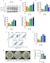
Results: Treatment with 160 μM Dox for 12 hours resulted in a reduction in CALR protein levels and the induction of apoptosis in HMC3 cells. The downregulation of CALR activated the IRE1/Caspase-12/Caspase-3 signaling pathway, leading to apoptosis. Similar apoptotic effects were observed in B. suis S2-infected HMC3 cells following Dox treatment.
Conclusion: Dox promotes apoptosis in B. suis S2-infected HMC3 cells by suppressing CALR expression and activating the IRE1/Caspase-12/Caspase-3 signaling pathway. These findings suggest that CALR regulation may serve as a potential therapeutic target for neurobrucellosis.
Keywords: Brucella suis S2 strain; HMC3; IRE1/Caspase-12/Caspase-3 pathway; apoptosis; doxycycline.
© 2025 Wang et al.
Conflict of interest statement
The authors declare that the research was conducted in the absence of any commercial or financial relationships that could be construed as a potential conflict of interest.
Figures


Similar articles
-
Doxycycline Induces Apoptosis of Brucella Suis S2 Strain-Infected HMC3 Microglial Cells by Activating Calreticulin-Dependent JNK/p53 Signaling Pathway.Front Cell Infect Microbiol. 2021 Apr 28;11:640847. doi: 10.3389/fcimb.2021.640847. eCollection 2021. Front Cell Infect Microbiol. 2021. PMID: 33996626 Free PMC article.
-
Brucella suis S2 strain inhibits IRE1/caspase-12/caspase-3 pathway-mediated apoptosis of microglia HMC3 by affecting the ubiquitination of CALR.mSphere. 2025 Mar 25;10(3):e0094124. doi: 10.1128/msphere.00941-24. Epub 2025 Feb 28. mSphere. 2025. PMID: 40019270 Free PMC article.
-
Brucella suis vaccine strain S2-infected immortalized caprine endometrial epithelial cell lines induce non-apoptotic ER-stress.Cell Stress Chaperones. 2015 May;20(3):399-409. doi: 10.1007/s12192-014-0564-x. Epub 2015 Jan 30. Cell Stress Chaperones. 2015. PMID: 25633898 Free PMC article.
-
Brucella suis Vaccine Strain 2 Induces Endoplasmic Reticulum Stress that Affects Intracellular Replication in Goat Trophoblast Cells In vitro.Front Cell Infect Microbiol. 2016 Feb 9;6:19. doi: 10.3389/fcimb.2016.00019. eCollection 2016. Front Cell Infect Microbiol. 2016. PMID: 26904517 Free PMC article.
-
[Mechanism of Gegen Qinlian Decoction in improving glucose metabolism in vitro and in vivo by alleviating hepatic endoplasmic reticulum stress].Zhongguo Zhong Yao Za Zhi. 2023 Oct;48(20):5565-5575. doi: 10.19540/j.cnki.cjcmm.20230516.401. Zhongguo Zhong Yao Za Zhi. 2023. PMID: 38114149 Chinese.
References
LinkOut - more resources
Full Text Sources
Research Materials
Miscellaneous

