. 2025 Apr 11:16:1589150.
doi: 10.3389/fphar.2025.1589150. eCollection 2025.

Ultrasensitive RNase H activity detection using the transcription-based hybrid probe and CRISPR/cas12a signal amplifier

Affiliations

Ultrasensitive RNase H activity detection using the transcription-based hybrid probe and CRISPR/cas12a signal amplifier

Sheng Ding et al. Front Pharmacol. .

Abstract

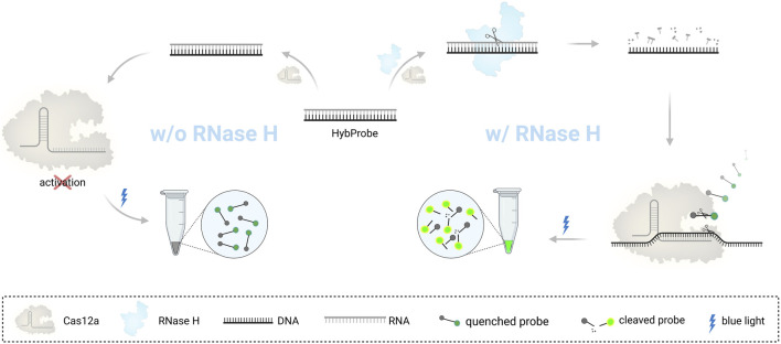
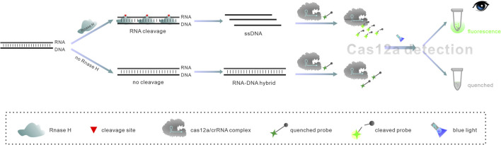
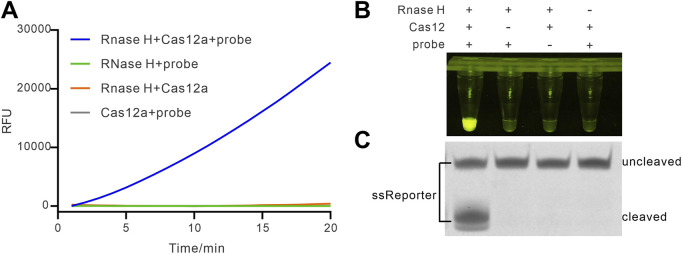
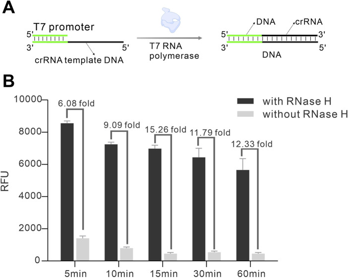
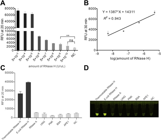
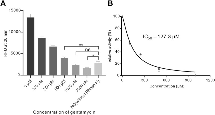
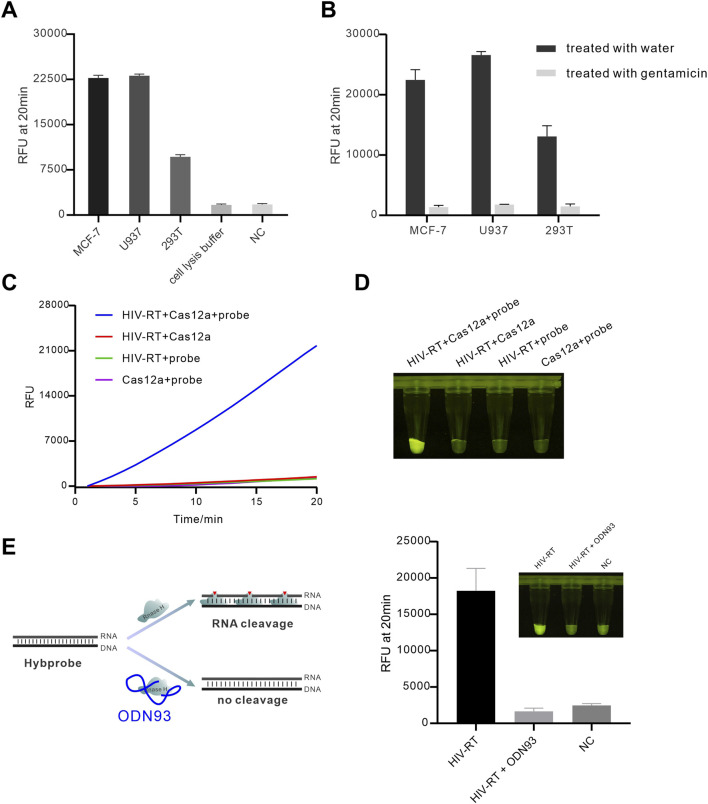
Ribonuclease H (RNase H), a critical functional protein in replication and genome stability, is emerging as a crucial therapeutic target for various diseases, including immune disorders. We present a transcription-based hybrid probe, referred to as Hybprobe, and a CRISPR/Cas12a signal amplifier for the rapid, sensitive, and low-cost detection of RNase H activity. In this method, the RNA strand of the Hybprobe is specifically cleaved by RNase H, releasing a single-stranded DNA activator that facilitates recognition and cleavage by the Cas12a/crRNA complex, triggering signal amplification via Cas12a's trans-cleavage activity. The proposed method demonstrates ultra-high sensitivity, capable of detecting RNase H as low as 9.02 × 10⁻¹⁰ U/μL, making it approximately 1,000 times more sensitive than several previously reported methods. Furthermore, we demonstrated the application of this method for RNase H inhibitor evaluation and its practical use across various biological samples, including cell extracts and HIV reverse transcriptase. In summary, the results suggest that this method is a promising tool for the highly sensitive detection of RNase H and the diagnosis of diseases associated with RNase H.

Keywords: CRISPR/Cas12a; Rnase H; activity detection; hybprobe; inhibitor screening.


Conflict of interest statement

The authors declare that the research was conducted in the absence of any commercial or financial relationships that could be construed as a potential conflict of interest.

Figures

None
Graphical abstract
FIGURE 1
FIGURE 1
Schematic diagram of the RNase H detection based on HybProbe.
FIGURE 2
FIGURE 2
Feasibility of HybProbe for APE1 detection. (A) Real-time fluorescence assay of CRISPR/Cas12a signal amplifier triggered by different reactions containing different components. (B) End-point fluorescence intensity difference between the reactions with different components. (C) Electrophoretic analysis of cleavage of ssReporter.
FIGURE 3
FIGURE 3
Optimization of the time for Hybprobe preparation and crRNA transcription. (A) Schematic of HybProbe preparation and crRNA transcription. (B) Investigation of the effect of different transcription times on the detection signal.
FIGURE 4
FIGURE 4
Investigation of the sensitivity and the specificity of the assay. (A) Endpoint fluorescence intensity at different RNase H concentrations. (B) The linear relationship between fluorescence intensity and the logarithm of RNase H concentration. **(p < 0.01) indicates a significant difference; ns indicates no significant difference (p > 0.05). (C) Detection of endpoint fluorescence intensity for different proteins. (D) Endpoint visualization results from Figure (C). The concentrations of thermostable RNase H, RNase H, RNase A, PNK, BSA, and APE1 used were 0.05 U/μL, 0.05 U/μL, 1 U, 100 μg/mL, 10 U, 1 mg/mL, and 10 U, respectively.
FIGURE 5
FIGURE 5
Inhibition assay of RNase H. (A) Investigation of the inhibition of RNase H (final concentration 1 × 10^-5 U/μL) by different concentrations of gentamicin; (B) Analysis of the inhibition efficiency of gentamicin. * (p < 0.05) and ** (p < 0.01) indicate significant differences; ns indicates no significant difference (p > 0.05).
FIGURE 6
FIGURE 6
Detecting of cellular and viral RNase H. (A) Real-time fluorescence curves for different cell extracts were obtained; (B) investigation of the inhibition of cellular RNase H. The cell lysates were derived from approximately 50,000 cells, and the gentamicin concentration was 1 mM; (C) Real-time fluorescence curves for detection of RNase H activity of HIV reverse transcriptase; (D) Endpoint fluorescence image for detection of RNase H activity of HIV reverse transcriptase. The used concentration of HIV-RT was 31 nM. (E) Inhibition of HIV-RT RNase H activity by aptamer ODN93. The concentration of ODN93 was 1 uM.

Similar articles

References

    1. Aden K., Bartsch K., Dahl J., Reijns M. A. M., Esser D., Sheibani-Tezerji R., et al. (2019). Epithelial RNase H2 maintains genome integrity and prevents intestinal tumorigenesis in mice. Gastroenterology 156 (1), 145–159. 10.1053/j.gastro.2018.09.047 - DOI - PMC - PubMed
    1. Andreola M.-L., Pileur F., Calmels C., Ventura M., Tarrago-Litvak L., Toulmé J.-J., et al. (2001). DNA aptamers selected against the HIV-1 RNase H display in vitro antiviral activity. Biochemistry 40 (34), 10087–10094. 10.1021/bi0108599 - DOI - PubMed
    1. Arraiano C. M., Andrade J. M., Domingues S., Guinote I. B., Malecki M., Matos R. G., et al. (2010). The critical role of RNA processing and degradation in the control of gene expression. FEMS Microbiol. Rev. 34 (5), 883–923. 10.1111/j.1574-6976.2010.00242.x - DOI - PubMed
    1. Bartsch K., Knittler K., Borowski C., Rudnik S., Damme M., Aden K., et al. (2017). Absence of RNase H2 triggers generation of immunogenic micronuclei removed by autophagy. Hum. Mol. Genet. 26 (20), 3960–3972. 10.1093/hmg/ddx283 - DOI - PubMed
    1. Bubeck D., Reijns M. A., Graham S. C., Astell K. R., Jones E. Y., Jackson A. P. (2011). PCNA directs type 2 RNase H activity on DNA replication and repair substrates. Nucleic Acids Res. 39 (9), 3652–3666. 10.1093/nar/gkq980 - DOI - PMC - PubMed

LinkOut - more resources