Solid Variant of Aneurysmal Bone Cyst (SVABC) of the Left Fibula Bone: A Rare Case Report
- PMID: 40290462
- PMCID: PMC12024465
- DOI: 10.2147/IMCRJ.S511228
Solid Variant of Aneurysmal Bone Cyst (SVABC) of the Left Fibula Bone: A Rare Case Report
Abstract
Introduction: Solid variant of aneurysmal bone cyst (SVABC) is a rare subtype of aneurysmal bone cyst (ABC) that presents as a solid, densely sclerotic lesion that can be more difficult to distinguish from other bone tumors and can lead to a wrong diagnosis. The SVABC rarely occurs in the long bones of the lower extremities.
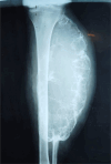
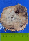
Case presentation: In this report, we present a rare case of SVABC in a 25-year-old male patient, which was seen in the left fibula bone. The patient had a history of trauma for 7 years. A physical examination showed a non-tender swelling in his left fibula bone. A preoperative frontal radiograph showed a huge expansile lytic lesion with trabeculations in the proximal 2/3 of the left fibula. Magnetic Resonance Imaging (MRI) showed fibula with multiple cystic areas in the lesion, some containing fluid-fluid levels. Excision of the mass was performed. Histopathological examination of the surgical specimen of the left fibula mass confirmed that the lesion was SVABC and showed largely solid proliferation of mildly pleomorphic oval to spindle cells with giant cells. A postoperative frontal radiograph of the leg demonstrated proximal 2/3 of the left fibulectomy with no lesion recurrence.
Conclusion: SVABC of the left fibula bone is a rare condition often misdiagnosed due to overlapping features with other aggressive bone lesions. Accurate diagnosis necessitates a multidisciplinary approach integrating clinical, imaging, and histopathology, with early surgical intervention being the gold standard for favorable outcomes. Surgeons must be cautious of postoperative complications like bleeding and neurological deficits, emphasizing the role of histopathology in preventing unnecessary surgeries. Future studies should focus on long-term follow-up and comparative treatment efficacy studies to enhance understanding and management of SVABC.
Keywords: SVABC; bone; excision; osteoclastic giant cells; radiology.
© 2025 Ahmed et al.
Conflict of interest statement
The authors declare no conflicts of interest in this work.
Figures


Similar articles
-
A Rare Subtype in an Uncommon Location: Multimodal Radiologic and Histopathologic Evaluation of a Solid Variant Aneurysmal Bone Cyst in the Radial Head.J Belg Soc Radiol. 2025 Jul 4;109(1):27. doi: 10.5334/jbsr.4012. eCollection 2025. J Belg Soc Radiol. 2025. PMID: 40620553 Free PMC article.
-
Solid Variant of Aneurysmal Bone Cyst Masquerading as Malignancy.J Clin Diagn Res. 2017 Jul;11(7):ED35-ED36. doi: 10.7860/JCDR/2017/25950.10306. Epub 2017 Jul 1. J Clin Diagn Res. 2017. PMID: 28892920 Free PMC article.
-
Solid variant of aneurysmal bone cyst of the heel: a case report.J Med Case Rep. 2011 Apr 12;5:145. doi: 10.1186/1752-1947-5-145. J Med Case Rep. 2011. PMID: 21486467 Free PMC article.
-
Atypical presentation of a solid-variant orbital aneurysmal bone cyst with a literature review.Neurochirurgie. 2018 Dec;64(6):431-433. doi: 10.1016/j.neuchi.2018.10.001. Epub 2018 Nov 6. Neurochirurgie. 2018. PMID: 30413280 Review.
-
Langerhans cell histiocytosis with aneurysmal bone cyst-like changes: a case-based literature review.Childs Nerv Syst. 2023 Nov;39(11):3057-3064. doi: 10.1007/s00381-023-06108-7. Epub 2023 Jul 31. Childs Nerv Syst. 2023. PMID: 37522932 Free PMC article. Review.
Cited by
-
A Rare Subtype in an Uncommon Location: Multimodal Radiologic and Histopathologic Evaluation of a Solid Variant Aneurysmal Bone Cyst in the Radial Head.J Belg Soc Radiol. 2025 Jul 4;109(1):27. doi: 10.5334/jbsr.4012. eCollection 2025. J Belg Soc Radiol. 2025. PMID: 40620553 Free PMC article.
References
Publication types
LinkOut - more resources
Full Text Sources

