. 2025 Apr 21:19:3059-3076.
doi: 10.2147/DDDT.S509046. eCollection 2025.

Gui-Pi-Tang Defers Skeletal Muscle and Cardiac Muscle Aging by Promoting Mitochondrial Remodeling

Affiliations

Gui-Pi-Tang Defers Skeletal Muscle and Cardiac Muscle Aging by Promoting Mitochondrial Remodeling

Changjiu Cai et al. Drug Des Devel Ther. .

Abstract

Purpose: To determine whether Gui-Pi-Tang (GPT) has protective effects on skeletal muscle and cardiac muscle in aged mice.

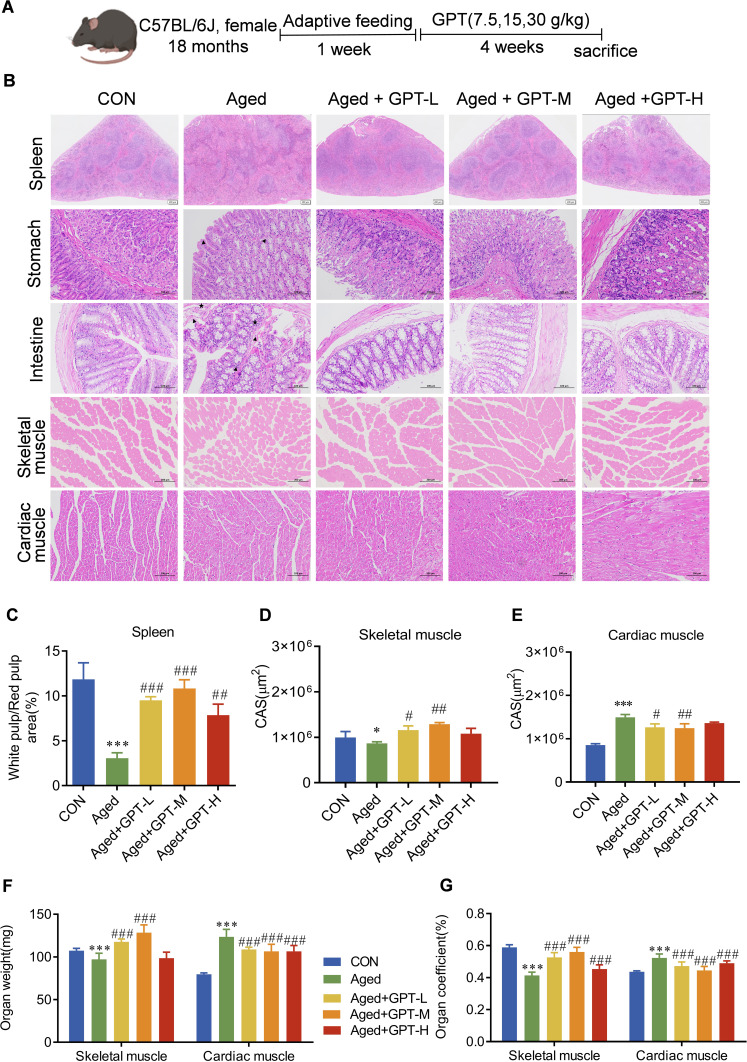
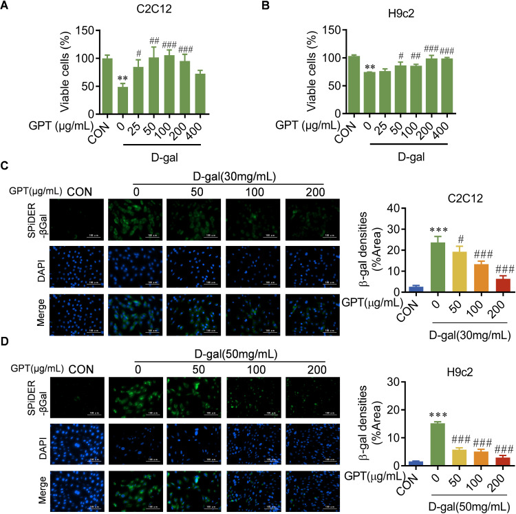
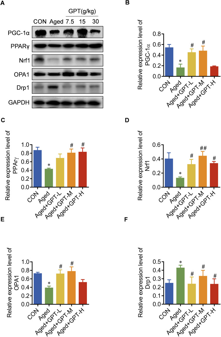
Methods: This study used C57BL6/J mice to establish an in vivo natural aging model, while D-galactose (D-gal)-injured C2C12 and H9c2 cells were employed to create in vitro aging cell models. Hematoxylin and eosin (H&E) staining was used to assess the effect of GPT on skeletal and cardiac muscle in aged mice. Protection against age-induced cellular damage by GPT was assessed in C2C12 and H9c2 cells using β-galactosidase staining. Mitochondrial morphology, structure, and function were analyzed using transmission electron microscopy, Seahorse assays, and ATP content measurements. Potential mechanisms by which GPT regulates mitochondrial homeostasis were examined using Western blot analysis.

Results: GPT treatment significantly improved the alignment of skeletal muscle fibers, reduced gaps, and increased the cross-sectional area (CSA) of skeletal muscle in aged mice. It also reduced the CSA of cardiac muscle fibers, alleviating cardiomyocyte hypertrophy. Mitochondrial morphology was restored, and GPT reduced D-gal-induced β-galactosidase elevation. Furthermore, GPT protected mitochondrial morphological and structural integrity in the skeletal and cardiac muscles of aged mice and improved mitochondrial respiratory function and ATP levels in D-gal-injured C2C12 and H9c2 cells. GPT treatment increased the levels of mitochondrion-associated proteins PGC-1α, PPARγ, Nrf1, and OPA1 in the skeletal and cardiac muscle of aged mice. Moreover, GPT modulated Drp1 expression, with increases in aged skeletal muscle and decreases in aged cardiac muscle.

Conclusion: These findings suggest that GPT helps maintain mitochondrial homeostasis by regulating mitochondrial remodeling, thereby alleviating skeletal and cardiac muscle damage in aged mice.

Keywords: Gui-Pi-Tang; mitochondrial homeostasis; myocardium; senescence; skeletal muscle.


Conflict of interest statement

The authors report no conflicts of interest in this work.

Figures

None
Graphical abstract
Figure 1
Figure 1
Total ion chromatograms for GPT obtained on a UHPLC-Q-Exactive HF platform in (A) positive and (B) negative ion modes.
Figure 2
Figure 2
Protective effects of GPT in skeletal and cardiac muscles of aged mice. (A) Schedule of animal experiments. (B) Morphological changes in spleen (scale bar = 100 μm), stomach (scale bar = 100 μm), intestine (scale bar = 100 μm), skeletal muscle (scale bar = 200 μm) and cardiac muscle (scale bar = 100 μm) were assessed by H&E staining. “▲” indicates infiltration of neutrophils and mononuclear inflammatory cells in the mucosa or submucosa. “★” indicates the degeneration or necrosis of mucosal epithelium. (C) White/red pulp area of the spleen. (D and E) CSA analysis of H&E staining for skeletal and cardiac muscles. (F) Organ weights of skeletal and cardiac muscles. (G) Organ coefficients of skeletal and cardiac muscles. Data represent means ± SD (n = 5). Compared with the control group: *P < 0.05, ***P < 0.001; compared with the model group: #P < 0.05, ##P < 0.01, ###P < 0.001.
Figure 3
Figure 3
Protective effects of GPT in D-gal-injured C2C12 and H9c2 cells. (A and B) MTT assays were used for measuring the effect of GPT on the survival of D-gal-injured C2C12 and H9c2 cells. (C and D) Representative images of SA-β-gal staining in D-gal injured C2C12 and H9c2 cells incubated with GTP for 48 h. Scale bar = 100 μm. Data (n = 5) represent means ± SD. Compared with the control group: **P < 0.01, ***P < 0.001; compared with the model group: #P < 0.05, ##P < 0.01, ###P < 0.001.
Figure 4
Figure 4
Effect of GPT on mitochondrial function in skeletal muscles of aged mice and D-gal injured C2C12 cells. (A) Ultrastructure (2,500×) and electron microscopy images (40,000×) of mitochondria in skeletal muscle. Mitochondrial area statistics (n = 5). (B) Seahorse curves of mitochondrial stress experiments in C2C12 cells. (C and D) Basal respiration and Maximal respiration of Seahorse curves. (E) ATP production in C2C12 cells. (F) Mitochondrial images (40,000×) and mitochondrial area statistics (n = 3) of C2C12 cells. Compared with control group, **P < 0.01, ***P < 0.001; compared with the model group: #P < 0.05, ##P < 0.01, ###P < 0.001.
Figure 5
Figure 5
Effect of GPT on mitochondrial function in cardiac muscle of aged mice and D-gal injured H9c2 cells. (A) Ultrastructure (2,500×) and electron microscopy images (40,000×) of mitochondria in cardiac muscle. Mitochondrial area statistics (n = 5). (B) Seahorse curves of mitochondrial stress experiments in H9c2 cells. (C and D) Basal respiration and Maximal respiration of Seahorse curves. (E) ATP production in H9c2 cells. (F) Mitochondrial images (40,000×) and mitochondrial area statistics (n = 3) of H9c2 cells. Compared with control group, *P < 0.05, ***P < 0.001; compared with the model group: #P < 0.05, ##P < 0.01.
Figure 6
Figure 6
The influence of GPT on mitochondrial remodeling in skeletal muscles of aged mice. (A) Protein levels of PGC-1α, PPARγ, OPA1, Drp1, and Nrf1 were evaluated by Western blot. (BF) Densitometry was used to quantify PGC-1α, PPARγ, OPA1, Drp1, and Nrf1 levels. Data (n=3) represent means ± SD. Compared with control group: *P < 0.05, **P < 0.01; compared with model group: #P < 0.05, ##P < 0.01.
Figure 7
Figure 7
The effects of GPT on mitochondrial remodeling in cardiac muscle of aged mice. (A) Protein expression of PGC-1α, PPARγ, OPA1, Drp1, and Nrf1 were evaluated by Western blot. (BF) Densitometry was used to quantify PGC-1α, PPARγ, OPA1, Drp1, and Nrf1 levels. Data (n=3) represent means ± SD. Compared with control group: *P < 0.05; compared with model group: #P < 0.05, ##P < 0.01.
Figure 8
Figure 8
GPT slows skeletal muscle and cardiac muscle aging by promoting mitochondrial remodeling.

References

    1. de Oliveira Zanuso B, de Oliveira Dos Santos AR, Miola VFB, Guissoni Campos LM, Spilla CSG, Barbalho SM. Panax ginseng and aging related disorders: a systematic review (1873-6815 (Electronic)). Exper Gerontol. 2022;161:111731. - PubMed
    1. Guo J, Huang X, Dou L, et al. Aging and aging-related diseases: from molecular mechanisms to interventions and treatments. Signal Transduct Target Ther. 2022;7(1):391. doi:10.1038/s41392-022-01251-0 - DOI - PMC - PubMed
    1. López-Otín C, Blasco MA, Partridge L, Serrano M, Kroemer G. Hallmarks of aging: an expanding universe. Cell. 2023;186(2):243–278. doi:10.1016/j.cell.2022.11.001 - DOI - PubMed
    1. Liu SZ, Marcinek DJ. Skeletal muscle bioenergetics in aging and heart failure. Heart Fail Rev. 2017;22(2):167–178. doi:10.1007/s10741-016-9586-z - DOI - PMC - PubMed
    1. Rosen RS, Yarmush ML. Current trends in anti-aging strategies. Annu Rev Biomed Eng. 2023;25(1):363–385. doi:10.1146/annurev-bioeng-120122-123054 - DOI - PubMed