

Spinal Metastases in Diffuse Intrinsic Pontine Glioma: A Rare Presentation of Rapid Neurological Decline
- PMID: 40291176
- PMCID: PMC12032994
- DOI: 10.7759/cureus.81289
Spinal Metastases in Diffuse Intrinsic Pontine Glioma: A Rare Presentation of Rapid Neurological Decline
Abstract
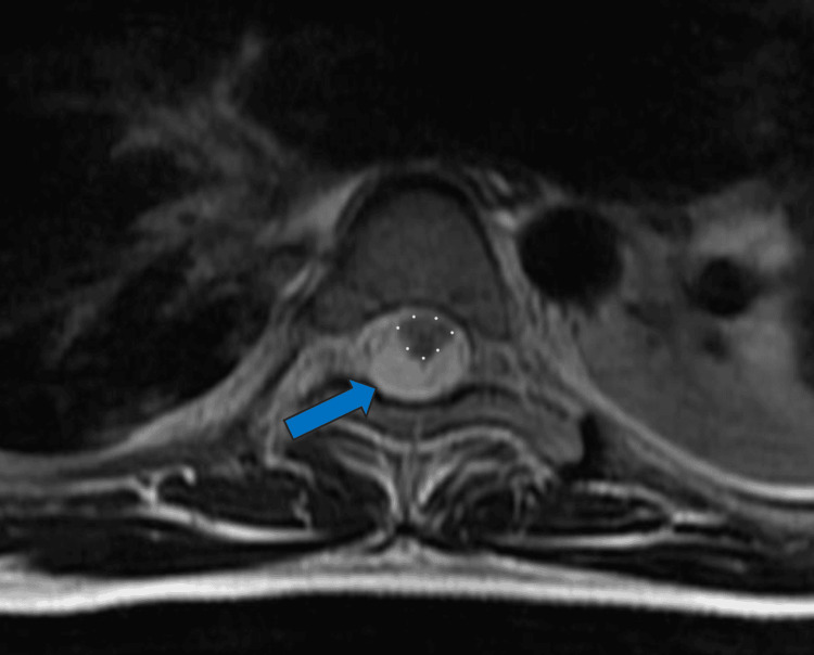
A nine-year-old male patient with a history of diffuse intrinsic pontine glioma (DIPG) presented with an episode of acute neurological deterioration approximately 18 months following completion of a primary course of radiation therapy to the brainstem for DIPG. His symptoms at that time included weakness in the lower extremities, urinary incontinence, and respiratory failure. Magnetic resonance imaging of the brain, cervical, thoracic, and lumbar spine revealed progression of the disease with metastatic deposits involving the craniospinal axis. He subsequently underwent a course of salvage radiation therapy to the craniospinal axis. This case highlights a rare but severe manifestation of metastatic DIPG with spinal involvement.
Keywords: brainstem tumor; conformal radiation therapy; craniospinal irradiation (csi); diffuse intrinsic pontine glioma; salvage radiation.
Copyright © 2025, Markey et al.
Conflict of interest statement
Human subjects: Consent for treatment and open access publication was obtained or waived by all participants in this study. Conflicts of interest: In compliance with the ICMJE uniform disclosure form, all authors declare the following: Payment/services info: All authors have declared that no financial support was received from any organization for the submitted work. Financial relationships: All authors have declared that they have no financial relationships at present or within the previous three years with any organizations that might have an interest in the submitted work. Other relationships: All authors have declared that there are no other relationships or activities that could appear to have influenced the submitted work.
Figures


References
-
- Pellot JE, De Jesus O. StatPearls [Internet] Treasure Island (FL): StatPearls Publishing; 2025. Diffuse intrinsic pontine glioma. - PubMed
-
- Leptomeningeal and subependymal seeding of diffuse intrinsic pontine glioma: a case report. Parenrengi MA, Prastikarunia R, Suryaningtyas W. Childs Nerv Syst. 2022;38:1643–1645. - PubMed
Publication types
LinkOut - more resources
Full Text Sources