

Diagnostic Pitfalls of External Auditory Canal Cholesteatoma: Insights From a Case Report
- PMID: 40291283
- PMCID: PMC12022382
- DOI: 10.7759/cureus.81200
Diagnostic Pitfalls of External Auditory Canal Cholesteatoma: Insights From a Case Report
Abstract
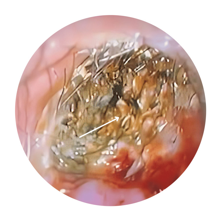
External auditory canal cholesteatoma (EACC) is a rare occurrence characterized by the keratinized mass of squamous epithelial cells in the external ear canal, leading to bone erosion and potential damage to surrounding structures. It's often misdiagnosed as otitis externa. Both conditions exhibit similar symptoms, which frequently result in misdiagnosis by clinicians. Though it is more common in older adults, EACC can occur in younger patients, as demonstrated in the case of an 18-year-old male. Insufficient diagnosis and delays in the management of this condition can result in significant complications. While the exact cause remains unclear, contributing factors may include canal trauma, chronic inflammation, or stenosis. We aim to share the experience of this pathology in order to highlight it as a differential diagnosis. This is particularly important in patients presenting with unresolved common ear symptoms.
Keywords: canal cholesteatoma; cholesteatoma; external auditory canal; external auditory canal cholesteatoma; otitis externa.
Copyright © 2025, Ramli et al.
Conflict of interest statement
Human subjects: Consent for treatment and open access publication was obtained or waived by all participants in this study. Conflicts of interest: In compliance with the ICMJE uniform disclosure form, all authors declare the following: Payment/services info: All authors have declared that no financial support was received from any organization for the submitted work. Financial relationships: All authors have declared that they have no financial relationships at present or within the previous three years with any organizations that might have an interest in the submitted work. Other relationships: All authors have declared that there are no other relationships or activities that could appear to have influenced the submitted work.
Figures

Similar articles
-
Spontaneous External Auditory Canal Cholesteatoma: Case Series and Review of Literature.Indian J Otolaryngol Head Neck Surg. 2020 Mar;72(1):86-91. doi: 10.1007/s12070-019-01755-2. Epub 2019 Oct 31. Indian J Otolaryngol Head Neck Surg. 2020. PMID: 32158662 Free PMC article.
-
External auditory canal cholesteatoma and benign necrotising otitis externa: clinical study of 95 cases in the Capital Region of Denmark.J Laryngol Otol. 2018 Jun;132(6):514-518. doi: 10.1017/S0022215118000750. Epub 2018 Jun 11. J Laryngol Otol. 2018. PMID: 29888691
-
[Burow's solution treatment for external auditory canal and mastoid cavity cholesteatoma].Nihon Jibiinkoka Gakkai Kaiho. 2010 Jun;113(6):549-55. doi: 10.3950/jibiinkoka.113.549. Nihon Jibiinkoka Gakkai Kaiho. 2010. PMID: 20653194 Japanese.
-
External Ear Disease: Keratinaceous Lesions of the External Auditory Canal.Otolaryngol Clin North Am. 2023 Oct;56(5):897-908. doi: 10.1016/j.otc.2023.06.013. Epub 2023 Aug 5. Otolaryngol Clin North Am. 2023. PMID: 37550109 Review.
-
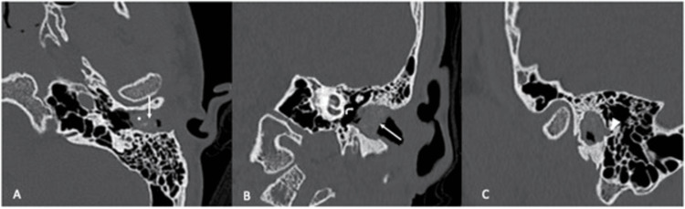
Computed Tomography Features of External Auditory Canal Cholesteatoma: A Pictorial Review.Curr Probl Diagn Radiol. 2015 Nov-Dec;44(6):511-6. doi: 10.1067/j.cpradiol.2015.05.001. Epub 2015 May 7. Curr Probl Diagn Radiol. 2015. PMID: 26050022 Review.
References
-
- Ear canal cholesteatoma: meta-analysis of clinical characteristics with update on classification, staging and treatment. Dubach P, Mantokoudis G, Caversaccio M. Curr Opin Otolaryngol Head Neck Surg. 2010;18:369–376. - PubMed
-
- Acquired ear canal cholesteatoma in congenital aural atresia/stenosis. Casale G, Nicholas BD, Kesser BW. Otol Neurotol. 2014;35:1474–1479. - PubMed
-
- Primary external auditory canal cholesteatoma of 301 ears: a single-center study. He G, Wei R, Chen L, et al. Eur Arch Otorhinolaryngol. 2022;279:1787–1794. - PubMed
-
- Cholesteatoma of external auditory canal: a case report. Sy A, Regonne EJ, Missie M, Ndiaye M. Pan Afri Med Jr. 2019
Publication types
LinkOut - more resources
Full Text Sources