

"Close to the tip, with little bone to grip": stabilizing two periprosthetic proximal femur fractures above a distal femur megaprosthesis using a combination of DHS and 3.5 mm screws
- PMID: 40291411
- PMCID: PMC12032374
- DOI: 10.1016/j.tcr.2025.101167
"Close to the tip, with little bone to grip": stabilizing two periprosthetic proximal femur fractures above a distal femur megaprosthesis using a combination of DHS and 3.5 mm screws
Abstract
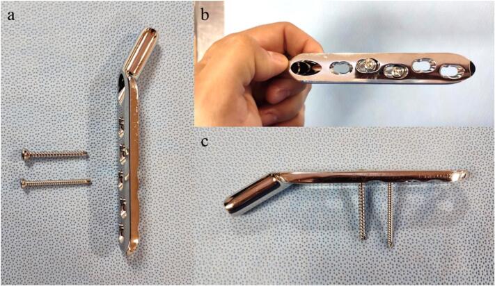
The incidence of periprosthetic fractures is increasing, presenting significant challenges due to patient longevity and the complexity of repeated surgeries. This report details the successful treatment of a previously unreported periprosthetic fracture pattern using a modified dynamic hip screw (DHS) technique. Two cases involved patients with extracapsular fractures in short proximal femur segments above megaprostheses. The fractures were reduced and stabilized with a DHS device, complemented by 3.5 mm screws from a different manufacturer to achieve effective bicortical fixation around the thick stems. Early weight-bearing was initiated postoperatively, with both patients achieving fracture healing without mechanical complications. This approach highlights the importance of careful preoperative planning and the selection of appropriate fixation methods, particularly in complex cases where traditional solutions may not be viable.
Keywords: Dynamic hip screw (DHS); Megaprosthesis; Periprosthetic fracture; Proximal femur.
© 2025 The Authors. Published by Elsevier Ltd.
Conflict of interest statement
The authors declare financial conflicts of interest with Smith & Nephew. Zimmer-Biomet, Stryker, Link Orthopaedics, Arthrex, and MBA Surgical Empowerment.
Figures


Similar articles
-
Revision arthroplasty in periprosthetic fractures of the proximal femur.Oper Orthop Traumatol. 2014 Oct;26(5):455-68. doi: 10.1007/s00064-014-0305-4. Epub 2014 Sep 28. Oper Orthop Traumatol. 2014. PMID: 25261284 Clinical Trial.
-
Total hip arthroplasty after failed fixation of a proximal femur fracture: Analysis of 59 cases of intra- and extra-capsular fractures.Orthop Traumatol Surg Res. 2018 Sep;104(5):681-686. doi: 10.1016/j.otsr.2018.04.015. Epub 2018 Jun 13. Orthop Traumatol Surg Res. 2018. PMID: 29908356
-
Dynamic Hip Screw Fixation Vs. Proximal Femur Nail For Unstable Per-Trochanteric Fractures: A Comparative Analysis Of Outcomes And Complications.J Ayub Med Coll Abbottabad. 2021 Jan-Mar;33(1):34-38. J Ayub Med Coll Abbottabad. 2021. PMID: 33774951
-
Non-neoplastic indications and outcomes of the proximal and distal femur megaprosthesis: a critical review.Knee Surg Relat Res. 2020 Apr 9;32(1):18. doi: 10.1186/s43019-020-00034-7. Knee Surg Relat Res. 2020. PMID: 32660578 Free PMC article. Review.
-
Comparison of young femoral neck fractures treated by femoral neck system, multiple cancellous screws and dynamic hip screws: a retrospectively comparison study.BMC Musculoskelet Disord. 2024 Mar 2;25(1):188. doi: 10.1186/s12891-024-07319-y. BMC Musculoskelet Disord. 2024. PMID: 38431562 Free PMC article. Review.
References
-
- Peiró J.V.A., Ruiz M.J., Hernández J.T., Serra J.T., Marsá J.S., Vázquez J.A.P., et al. The inverted Vancouver C fracture. Case series of unstable proximal femur fractures above a knee revision stem treated by short cephalomedullary nail and lateral submuscular overlapping plate. Eur. J. Orthop. Surg. Traumatol. 2021;31:193–198. doi: 10.1007/s00590-020-02738-8. - DOI - PubMed
-
- Gonzalez-Morgado D., Andres-Peiro J.V., Selga Marsa J., Alberto Piedra Calle C., Francesc Nomdedeu Sancho J., Teixidor Serra J., et al. Open reduction and polyaxial plating for stemmed knee periprosthetic fractures: a case series. SICOT J. 2023;9:24. doi: 10.1051/sicotj/2023022. - DOI - PMC - PubMed
Publication types
LinkOut - more resources
Full Text Sources
