. 2025 Apr 21:18:5383-5398.
doi: 10.2147/JIR.S496387. eCollection 2025.

IL-22 Alleviates Sepsis-Induced Acute Lung Injury by Inhibiting Epithelial Cell Apoptosis Associated with STAT3 Signalling

Affiliations

IL-22 Alleviates Sepsis-Induced Acute Lung Injury by Inhibiting Epithelial Cell Apoptosis Associated with STAT3 Signalling

Chiying Zhu et al. J Inflamm Res. .

Abstract

Purpose: Sepsis is a critical condition characterized by organ dysfunction due to an aberrant response to infection, which results in a life-threatening situation. The lung, which is the most vulnerable target organ, is often severely damaged during sepsis. Research has demonstrated that interleukin-22 (IL-22), which is secreted by various immunocytes, can mitigate inflammation-associated diseases. Nevertheless, the precise function of IL-22 in sepsis-induced acute lung injury (SALI) is still unclear. This study aimed to investigate the therapeutic efficacy of IL-22 in sepsis and explore the regulatory mechanisms involved.

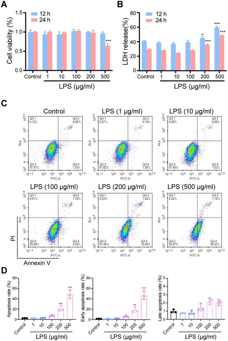
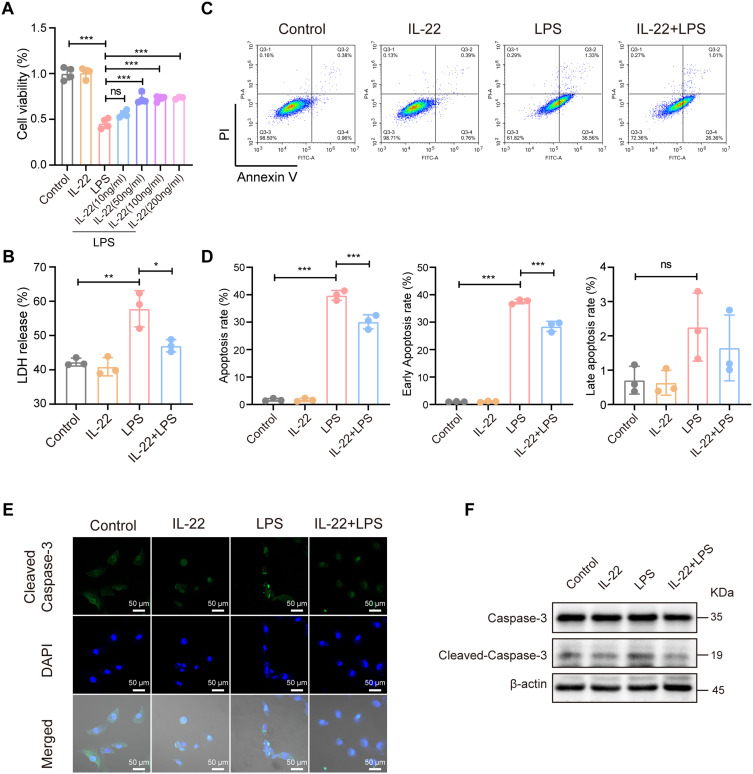
Methods: A mouse caecal ligation and puncture (CLP) model of sepsis was established, and the effect of IL-22 was investigated as indicated. Immunohistochemistry, qRT‒PCR, ELISA, immunofluorescence, TUNEL, Western blotting, and flow cytometry assays were applied to investigate the protective efficacy and involved pathways. Additionally, an in vitro model of lipopolysaccharide (LPS)-induced bronchial epithelial cell (BEAS-2B) apoptosis was established, and these cells were treated with or without recombinant IL-22 (rIL-22) to further evaluate the effect of IL-22 and the underlying mechanism.

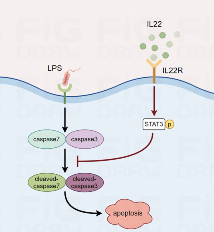
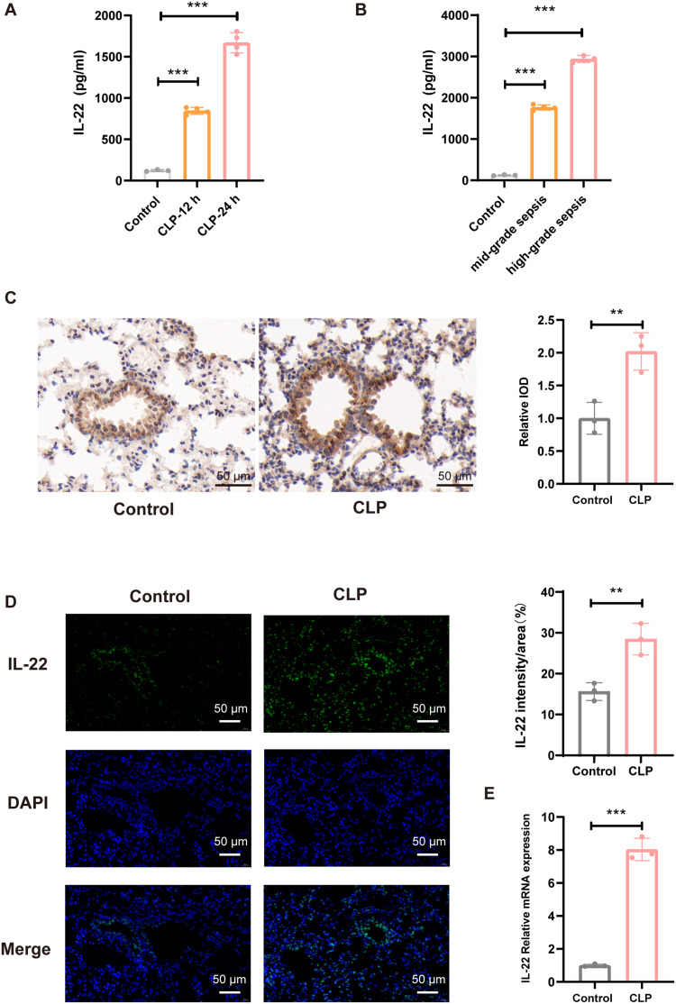
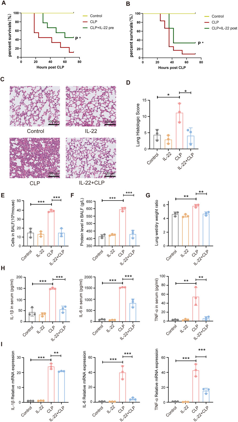
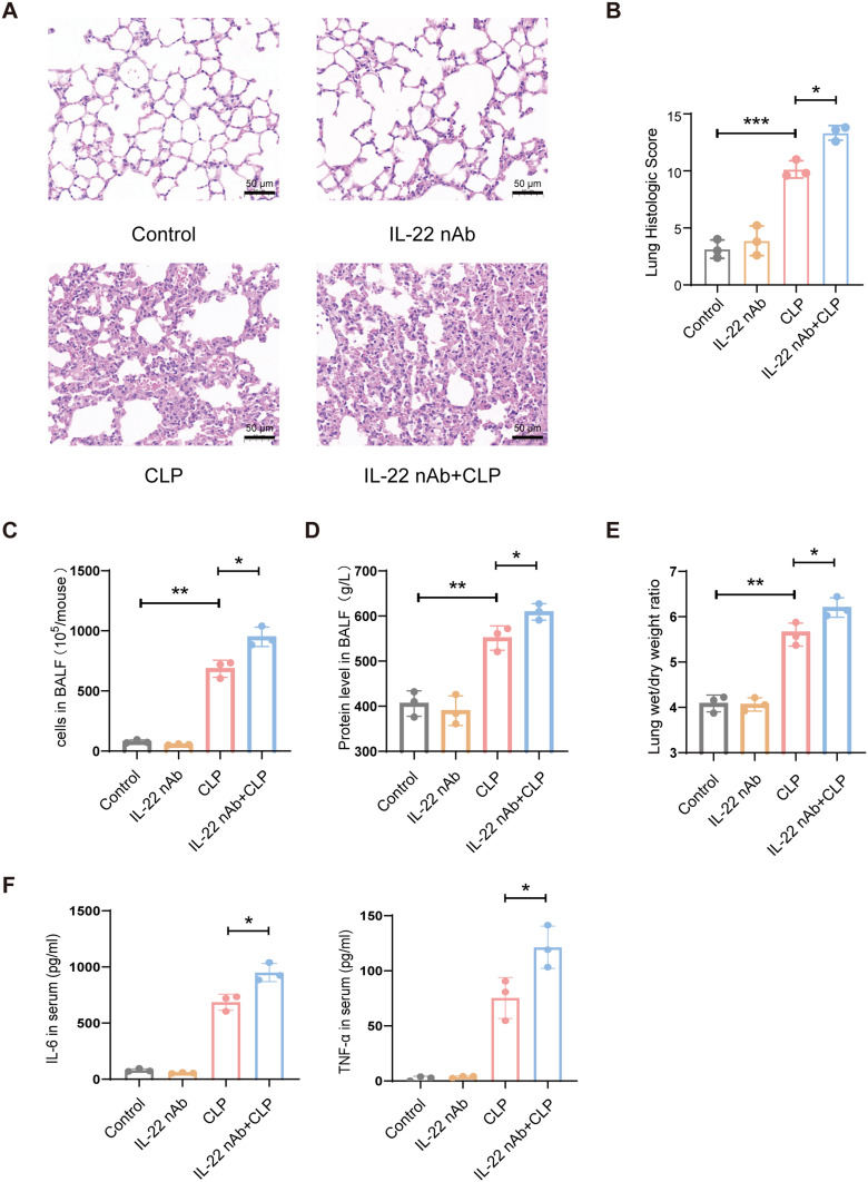
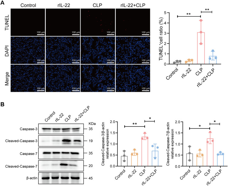
Results: The experimental results clearly confirmed that the levels of IL-22 were increased in the serum and lung tissue after CLP. The administration of rIL-22 was observed to increase the survival rate of septic mice. Notably, rIL-22 treatment resulted in decreased levels of proteins and a decreased cell number in the bronchoalveolar lavage fluid, as well as in a reduction in inflammatory cytokine release into the serum. Importantly, rIL-22 mitigated SALI by inhibiting lung cell apoptosis in septic mice. Furthermore, the results revealed that rIL-22 attenuated apoptosis of lung epithelial cells via the activation of the STAT3 signalling pathway.

Conclusion: The results of this study suggest that IL-22 alleviates lung epithelial cell apoptosis to protect mice against SALI in association with the STAT3 signalling pathway, highlighting the potential therapeutic value of IL-22 against sepsis.

Keywords: IL-22; STAT3; acute lung injury; apoptosis; sepsis.


Conflict of interest statement

The authors report no conflicts of interest in this work. This paper has been uploaded as a preprint to the Research Square website: https://www.researchsquare.com/article/rs-4198943/v1.

Figures

None
Graphical abstract
Figure 1
Figure 1
The expression of IL-22 was increased in the serum and lungs of septic mice. The mice were divided into a control group (Control) and a CLP group (CLP). Lung tissue was collected 12 hours after CLP at the indicated times (n=3). (A) Serum levels of IL-22 in mice at different time points after CLP. (B) Serum levels of IL-22 in mice subjected to different grades of CLP at 24 hours. (C) Immunohistochemical staining for IL-22 in lung sections. Reddish-brown staining indicates IL-22-positive cells. Scale bar, 50 μm. (D) IL-22 in lung sections was stained for immunofluorescence imaging. Green signals are from immunostaining with an IL-22 antibody, and blue signals represent nuclei that are stained with DAPI. Scale bar, 50 μm. (E) The mRNA level of IL-22 in lung tissue. Significant differences were determined by a two-tailed unpaired Student’s t-test (CE) or one-way ANOVA with Bonferroni’s multiple comparisons test (A and B). **P<0.01, ***P<0.001.
Figure 2
Figure 2
rIL-22 protects mice against SALI. (A) Survival of mice injected with rIL-22 or vehicle 1 hour before CLP surgery (n = 12). (B) Survival of mice injected with IL-22 or vehicle 1 hour after CLP surgery (n=9). (C) Lung sections were stained with H&E 12 hours after CLP, and the (D) lung injury score was calculated. Scale bar, 100 μm. (E) Total number of cells in the BALF. (F) Total protein content in the BALF. (G) Wet/dry weight ratios of the mouse lung tissue. (H) Serum IL-1β, IL-6 and TNF-α levels. (I) IL-1β, IL-6 and TNF-α mRNA levels in the lung tissue. Significant differences were determined by a log-rank (Mantel‒Cox) test (A and B) or one-way ANOVA with Bonferroni’s multiple comparisons test (DI). *P<0.05, **P<0.01, ***P<0.001.
Figure 3
Figure 3
IL-22 nAb exacerbates SALI. The mice were intraperitoneally injected with an IL-22 nAb or an equivalent amount of isotype IgG before CLP surgery. (A) Lung sections were stained with H&E 12 hours after CLP, and the (B) lung injury score was calculated. (C) Total number of cells in BALF. Scale bar, 50 μm. (D) Total protein content in the BALF. (E) Wet/dry weight ratios of mouse lungs. (F) Serum IL-6 and TNF-α levels. Significant differences were determined by one-way ANOVA with Bonferroni’s multiple comparisons test (BF). *P<0.05, **P<0.01, ***P<0.001.
Figure 4
Figure 4
IL-22 inhibits apoptosis in the lung tissue of septic mice. The mice were pretreated with rIL-22 and the lung tissue was collected at 12 hours after CLP (n=3). (A) TUNEL assays revealed that IL-22 reversed the rate of apoptosis in the lungs of mice after CLP. Scale bar, 100 μm. (B) Western blotting revealed that rIL-22 reversed the changes in the levels of cleaved caspase-3 and cleaved caspase-7 in septic mice. Significant differences were determined by one-way ANOVA with Bonferroni’s multiple comparisons test (A and B). *P<0.05, **P<0.01.
Figure 5
Figure 5
LPS promotes apoptosis in lung epithelial cells. (A) A CCK8 assay was used to examine the cytotoxicity of different concentrations of LPS at 12 hours and 24 hours. (B) Analysis of LDH levels in the cell culture medium after treatment with different concentrations of LPS for 12 hours and 24 hours. (C) BEAS-2B cells were stained with annexin V-FITC and propidium iodide (PI) and subjected to flow cytometry to examine the rates of apoptosis after treatment with different concentrations of LPS for 24 hours. (D) The percentages of early, late, and total apoptotic cells were quantified after LPS stimulation. All the samples were compared with the control. Significant differences were determined by one-way ANOVA with Bonferroni’s multiple comparisons test (A, B and D). *P<0.05, **P<0.01, ***P<0.001.
Figure 6
Figure 6
IL-22 attenuates LPS-induced apoptosis in BEAS-2B cells. (A) A CCK-8 assay was used to examine cytotoxicity in response to LPS stimulation with or without pretreatment with different concentrations of IL-22 for 24 hours. (B) Analysis of LDH release in response to LPS stimulation with or without IL-22 pretreatment for 24 hours. (C) BEAS-2B cells were stained with Annexin V-FITC and propidium iodide (PI) and subjected to flow cytometry to examine apoptosis in response to LPS stimulation with or without IL-22 pretreatment. (D) The percentages of early, late, and total apoptotic cells were quantified after LPS stimulation with or without IL-22 pretreatment. (E) The expression of cleaved caspase-3 in lung epithelial cells was examined via an immunofluorescence assay. Scale bar, 50 μm. (F) The expression of Caspase-3 and cleaved Caspase-3 in BEAS-2B cells in response to LPS stimulation and pretreatment with or without IL-22 for 24 hours was determined by Western blotting. Significant differences were determined by one-way ANOVA with Bonferroni’s multiple comparisons test (A, B and D). *P<0.05, **P<0.01, ***P<0.001.
Figure 7
Figure 7
IL-22 inhibits apoptosis by activating STAT3 signalling in BEAS-2B cells. (A) Western blotting analysis of STAT3 and p-STAT3 expression in BEAS-2B cells with or without rIL-22 pretreatment and LPS stimulation. (B) BEAS-2B cells were stained with annexin V-FITC and propidium iodide (PI) and subjected to flow cytometry to examine the apoptosis rates in response to LPS stimulation with or without IL-22 pretreatment and a STAT3 inhibitor (50 μM, NSC74859, MCE). The percentages of total apoptotic cells were quantified. Significant differences were determined by one-way ANOVA with Bonferroni’s multiple comparisons test. *P<0.05, ***P<0.001.

Similar articles

Cited by

References

    1. Rehn M, Chew MS, Olkkola KT, Ingi Sigurðsson M, Yli‐Hankala A, Hylander Møller M. Surviving sepsis campaign: international guidelines for management of sepsis and septic shock in adults 2021‐endorsement by the Scandinavian society of anaesthesiology and intensive care medicine. Acta Anaesthesiol Scand. 2022;66(5):634–635. doi:10.1111/aas.14045 - DOI - PMC - PubMed
    1. Gupta N, Matthay MA. Sepsis and stretch: synergistic effects on alveolar epithelial cell death? Crit Care Med. 2006;34(6):1846–1847. doi:10.1097/01.CCM.0000219378.52859.B1 - DOI - PubMed
    1. Kumar V. Pulmonary innate immune response determines the outcome of inflammation during pneumonia and sepsis-associated acute lung injury. Front Immunol. 2020;11:1722. doi:10.3389/fimmu.2020.01722 - DOI - PMC - PubMed
    1. Lam TY, Nguyen N, Peh HY, et al. ISM1 protects lung homeostasis via cell-surface GRP78-mediated alveolar macrophage apoptosis. Proc Natl Acad Sci U S A. 2022;119(4):e2019161119. doi:10.1073/pnas.2019161119 - DOI - PMC - PubMed
    1. Rutz S, Noubade R, Eidenschenk C, et al. Transcription factor c-Maf mediates the TGF-β-dependent suppression of IL-22 production in TH17 cells. Nat Immunol. 2011;12(12):1238–1245. doi:10.1038/ni.2134 - DOI - PubMed

LinkOut - more resources