Review
. 2025 Apr 11:16:1548194.
doi: 10.3389/fphys.2025.1548194. eCollection 2025.

Mechanisms underlying targeted mitochondrial therapy for programmed cardiac cell death

Affiliations
Review

Mechanisms underlying targeted mitochondrial therapy for programmed cardiac cell death

Fengting Jing et al. Front Physiol. .

Abstract

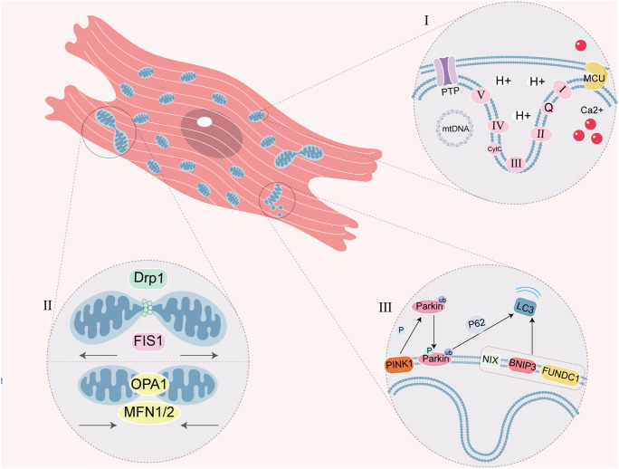
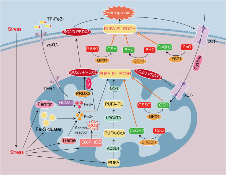
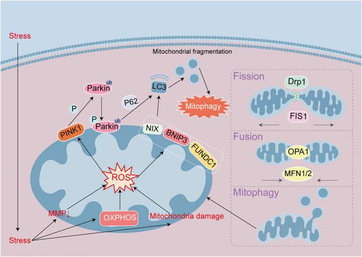
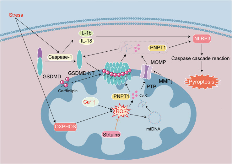
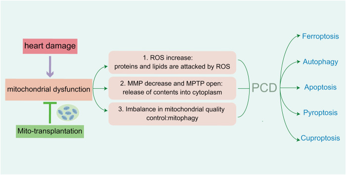
Heart diseases are common clinical diseases, such as cardiac fibrosis, heart failure, hypertension and arrhythmia. Globally, the incidence rate and mortality of heart diseases are increasing by years. The main mechanism of heart disease is related to the cellular state. Mitochondrion is the organ of cellular energy supply, participating in various signal transduction pathways and playing a vital role in the occurrence and development of heart disease. This review summarizes the cell death patterns and molecular mechanisms associated with heart disease and mitochondrial dysfunction.

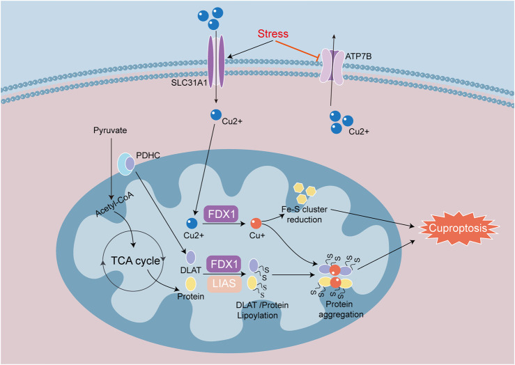
Keywords: PCD; apoptosis; autophagy; cardiomyocyte; cuproptosis; ferroptosis; mitochondria; pyroptosis.


Conflict of interest statement

The authors declare that the research was conducted in the absence of any commercial or financial relationships that could be construed as a potential conflict of interest.

Figures

FIGURE 1
FIGURE 1
Schematic diagram of myocardiocyte mitochondrial function. (I) ETC is a series of complexes used for mitochondrial respiratory function. PTP closes in physiological conditions and prohibits macromolecule shuttle, but it mediates the formation of MPTP in mitochondrial dysfunction. MCU is one of Ca2+ single transporter to regulate the Ca2+ concentration in the mitochondria, which Ca2+ overload cause MPTP. Circular mtDNA mainly encodes the protein of mitochondrial OXPHOS. (Ⅱ) Mitochondrial fission and fusion is a link of mitochondrial quality control. DRP1 and FIS1 regulate fission, OPA1 and MFN1/2 regulate fusion. (Ⅲ) Mitophagy is a physiological process, which mediated by ubiquitin ligase Parkin and PINK1 or mitophagy receptor (NIX, BNIP3 and FUNDC1). P62 is the ubiquitin-binding protein that promotes Parkin-dependent mitophagy.
FIGURE 2
FIGURE 2
Signaling mechanisms leading to ferroptosis in heart disease. Ferroptosis is caused by the accumulation of iron ions and lipid peroxides, and the destruction of the balance of the antioxidant system. Iron deposition is mainly caused by TF-Fe3+ entry into cells through TFR1, and the autophagic breakdown of Ferritin, Heme, and Fe-S cluster (NCOA4 mediates ferritin autophagy). Lipid peroxide deposition is due to the Fenton reaction between Fe2+ and ROS to form HO . PUFA converted to PL-PUFA-OOH via ACSL4, LPCAT3 and LOX with HO , and PL-PUFA-OOH accumulate on the cell membrane make ferroptosis. The antioxidant system GSH/GSSG/GPX4, CoQH2/CoQ/DHODH, CoQH2/CoQ/FSP1 and BH4/BH2/GCH1 prevent the generation of PUFA-PL-POOH. PRDX3 is an antioxidant enzyme in mitochondria, but it is attacked and formed SO2/3-PRDX3 by HO . SO2/3-PRDX3 is a specific marker of ferroptosis, and it suppresses xCT-cysteine uptake, reduces the antioxidant capacity of GPX4 and GSH/GSSG, and promotes ferroptosis.
FIGURE 3
FIGURE 3
Signaling mechanisms leading to autophagy in heart disease. Autophagy maintain a stable intracellular environment. Mitophagy initiation is through the PINK1/Parkin pathway or the autophagy receptor NIX/BNIP3/FUNDC1 pathway, respectively. Parkin and NIX/BNIP3/FUNDC1 can bind to LC3 proteins to mediate autophagy. Mitochondrial damage leads to MMP decline and ROS accumulation, and mitophagy initiates. Mitochondria maintain stability through division and fusion mediated by DRP1, FIS1, OPA1 and MFN.
FIGURE 4
FIGURE 4
Signaling mechanisms that lead to apoptosis in heart disease. Apoptosis is a process of cell death regulated by a series of genes. Endogenous apoptotic signaling is executed and regulated by the Bcl-2 family of proteins. They are divided into anti-apoptotic proteins Bcl-2 and Bcl-xL, and pro-apoptotic proteins Bak, Bid and Bax. Antiapoptotic proteins can inhibit the release of Cyt c. Apoptotic signals promote the dimer formation of proapoptotic proteins, which lead to form transmembrane pores in the mitochondrial surface. The integrity of the mitochondrial outer membrane is broken by decreasing MMP, changes in MOMP and open PTP channels. Cyt c released into the cytoplasm binds to Apaf1 to form Apoptosome, which activates Caspase 3 to mediate apoptosis. Mitochondrial damage leads to increase ROS and mtDNA damage. The release of Cyt c and mtDNA into the cytosol exacerbates cell apoptosis.
FIGURE 5
FIGURE 5
Signaling mechanisms leading to pyroptosis in heart disease. Pyroptosis is one of cell death induced by inflammatory effect. Stress activates Caspase-1, GSDMD, IL-1β and IL-18. The active fragment GSDMD-NT binds cardiolipin on the membrane to form a special pore, and MOMP causes MMP decrease and PTP channel opening. They cause the loss of cellular integrity and the release of cellular contents such as ROS, Cyt c, mtDNA, PNPT 1. OXPHOS and Ca2+ overload caused increased ROS to exacerbate mtDNA damage. Release of the exonuclease PNPT1 degrades the cytoplasmic mRNA, amplifying the downstream inflammatory response and aggravating pyroptosis. Sirtuin 5 reduce ROS and stabilize the mitochondrial environment.
FIGURE 6
FIGURE 6
Signaling mechanisms of cuproptosis in heart disease. Cuproptosis is a process of cell death mainly caused by the reduction of Fe-S cluster proteins and the aggregation of lipoylated proteins. SLC31A1 and ATP7B regulate intracellular copper in and out. Iron redox protein 1 (FDX 1) and fatty acid synthase (LIAS) are involved in the regulation of thiylation of DLAT and TCA cycle proteins. FDX1 converts Cu2+ to Cu+, suppresses the synthesis of Fe-S cluster proteins and induces cuproptosis. When Cu+ overaccumulates in mitochondria, Cu+ will bind to lipoylated proteins and induce heteromerization of lipoylated proteins, causing proteotoxic response to cuproptosis.
FIGURE 7
FIGURE 7
Injury of cardiomyocytes cause mitochondrial dysfunction, which mediate five PCDs in heart damage. The increasing of ROS attack related proteins and lipids. Abnormal MMP and channel-open release the contents of mitochondria. Imbalance of mitochondrial quality control lead to mitochondrial autophagy. Reducing the above aspects can alleviate mitochondrial dysfunction, as well as PCD-mediated myocardial injury.

Similar articles

References

    1. Baechler B. L., Bloemberg D., Quadrilatero J. (2019). Mitophagy regulates mitochondrial network signaling, oxidative stress, and apoptosis during myoblast differentiation. Autophagy 15 (9), 1606–1619. 10.1080/15548627.2019.1591672 - DOI - PMC - PubMed
    1. Balakrishnan M., Kenworthy A. K. (2024). Lipid peroxidation drives liquid-liquid phase separation and disrupts raft protein partitioning in biological membranes. J. Am. Chem. Soc. 146 (2), 1374–1387. 10.1021/jacs.3c10132 - DOI - PMC - PubMed
    1. Bersuker K., Hendricks J. M., Li Z., Magtanong L., Ford B., Tang P. H., et al. (2019). The CoQ oxidoreductase FSP1 acts parallel to GPX4 to inhibit ferroptosis. Nature 575 (7784), 688–692. 10.1038/s41586-019-1705-2 - DOI - PMC - PubMed
    1. Bi Y., Liu S., Qin X., Abudureyimu M., Wang L., Zou R., et al. (2024). FUNDC1 interacts with GPx4 to govern hepatic ferroptosis and fibrotic injury through a mitophagy-dependent manner. J. Adv. Res. 55, 45–60. 10.1016/j.jare.2023.02.012 - DOI - PMC - PubMed
    1. Cai W., Liu L., Shi X., Liu Y., Wang J., Fang X., et al. (2023). Alox15/15-HpETE aggravates myocardial ischemia-reperfusion injury by promoting cardiomyocyte ferroptosis. Circulation 147 (19), 1444–1460. 10.1161/circulationaha.122.060257 - DOI - PubMed

LinkOut - more resources