Comparative Study
. 2024 Oct 31;79(5):1047-1056.
doi: 10.32592/ARI.2024.79.5.1047. eCollection 2024 Oct.

Comparison of Sensitivities and Specificities of ELISA and Histopathology to Diagnose Feline Infectious Peritonitis

Affiliations
Comparative Study

Comparison of Sensitivities and Specificities of ELISA and Histopathology to Diagnose Feline Infectious Peritonitis

S Saeedi et al. Arch Razi Inst. .

Abstract

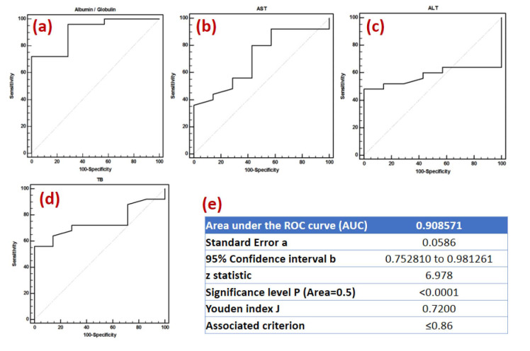
Feline infectious peritonitis (FIP) is one of the most prevalent viral infectious diseases in cats. It presents a number of challenges for veterinarians in terms of diagnosis. The objective of this study was to compare the sensitivity and specificity of ELISA with that of histopathology. Samples were obtained from 28 cats exhibiting signs consistent with feline infectious peritonitis (FIP) at the northwest animal clinics in Tehran, Iran, between January 2013 and 2015. Of the cats examined, five were deemed healthy, 14 exhibited indications of wet FIP, and nine displayed symptoms of dry FIP. Furthermore, the sensitivities and specificities of biochemical parameters were determined. The sensitivity and specificity of the ELISA test for diagnosing effusive FIP were found to be 100%, which was identical to the results obtained from histopathology. The AST (AUC=0.708) and total bilirubin (AUC=0.74) demonstrated moderate clinical accuracy in diagnosing FIP. The optical densities (ODs) in positive cats and the negative control group exhibited no statistically significant difference between the effusive and non-effusive forms of FIP. The Youden index was employed to determine the optimal cut-off point for the ratio of ODs in positive and negative cats, which was estimated to be 3.375. In conclusion, the ELISA demonstrated high predictive values for the diagnosis of effusive FIP and has the potential for use in the serological diagnosis of feline coronavirus infection.

Keywords: ELISA Test; Effusion; Feline infectious peritonitis; Histopathology.


Conflict of interest statement

None.

Figures

Figure 1
Figure 1
a) Pyogranulomatous granulomas are evident in the liver parenchyma. The arrow indicates the presence of neutrophils in proximity to the liver vein, indicative of vasculitis. H&E staining (×60), b) Liver, Pyogranulomatous granulomas, H&E staining (×100), and c) Liver. Arrow A indicates subcapsular fibrinous exudate, while arrow B illustrates the infiltration of neutrophils and mononuclear inflammatory cells (lymphocytes, plasma cells, and macrophages). H&E staining (×60).
Figure 2
Figure 2
a) The kidney tissue displays mononuclear inflammatory cells in the interstitial space (arrow A) and hyaline casts in the tubules (arrow B), indicating nephritis, a chronic form of feline infectious peritonitis lesion. H&E staining (×60), b) The Bowman's capsule and the wall of the blood vessels in glomeruli exhibited thickening, accompanied by the proliferation of mesenchymal cells in the glomeruli and the attachment of glomeruli epithelia to the walls of Bowman's capsules (synechia), indicative of membranoproliferative glomerulonephritis (MPGN). H&E staining (×60), and c) Synechia in membranoproliferative glomerulonephritis. H&E staining (×100).
Figure 3
Figure 3
a) A depletion of the Malphigian corpuscles of white pulps is accompanied by an infiltration of neutrophils in the red pulp space, which is indicative of feline infectious peritonitis lesions. H&E staining (×20) and b) depletion of the Malphigian corpuscles of white pulps. H & E staining (×60).
Figure 4
Figure 4
The presence of neutrophilic and mononuclear inflammatory cells among the myocytes (myocarditis) is indicative of feline infectious peritonitis lesions. Histological examination using the hematoxylin and eosin (H&E) staining method at a magnification of 100x.
Figure 5
Figure 5
a) albumin/globulin cut-off point, b) aspartate aminotransferase (AST), c) alanine aminotransferase (ALT), d) total bilirubin, and e) area under the ROC curve (AUC) and Youden index.

References

    1. Olival KJ, Hosseini PR, Zambrana-Torrelio C, Ross N, Bogich TL, Daszak P. Host and viral traits predict zoonotic spillover from mammals. Nature. 2017;546:646–650. - PMC - PubMed
    1. Nova N. Cross-Species Transmission of Coronaviruses in Humans and Domestic Mammals, What Are the Ecological Mechanisms Driving Transmission, Spillover, and Disease Emergence? Front Public Health. 2021;9:717941. - PMC - PubMed
    1. Mohammed Ibrahim O, Bara Allawe A, Ali Kadhim H. Isolation and Molecular Detection of Feline Infectious Peritonitis Virus. Archives Razi Institute. 2022;77(5):1709–1714. - PMC - PubMed
    1. Gao YY, Wang Q, Liang XY, Zhang S, Bao D, Zhao H, et al. An updated review of feline coronavirus: mind the two biotypes. Virus Research. 2023;326:199059. - PMC - PubMed
    1. Felten S, Hartmann K. Diagnosis of feline infectious peritonitis: a review of the current literature. Viruses. 2019;11(11):1068. - PMC - PubMed

Publication types

LinkOut - more resources