. 2025 Jun 10;93(6):e0008125.
doi: 10.1128/iai.00081-25. Epub 2025 Apr 28.

Evidence for a model of conformational change by the Plasmodium falciparum circumsporozoite protein during sporozoite development in the mosquito host through the use of camelid single-domain antibodies

Affiliations

Evidence for a model of conformational change by the Plasmodium falciparum circumsporozoite protein during sporozoite development in the mosquito host through the use of camelid single-domain antibodies

Rob Geens et al. Infect Immun. .

Abstract

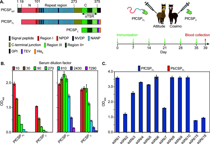
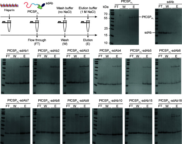
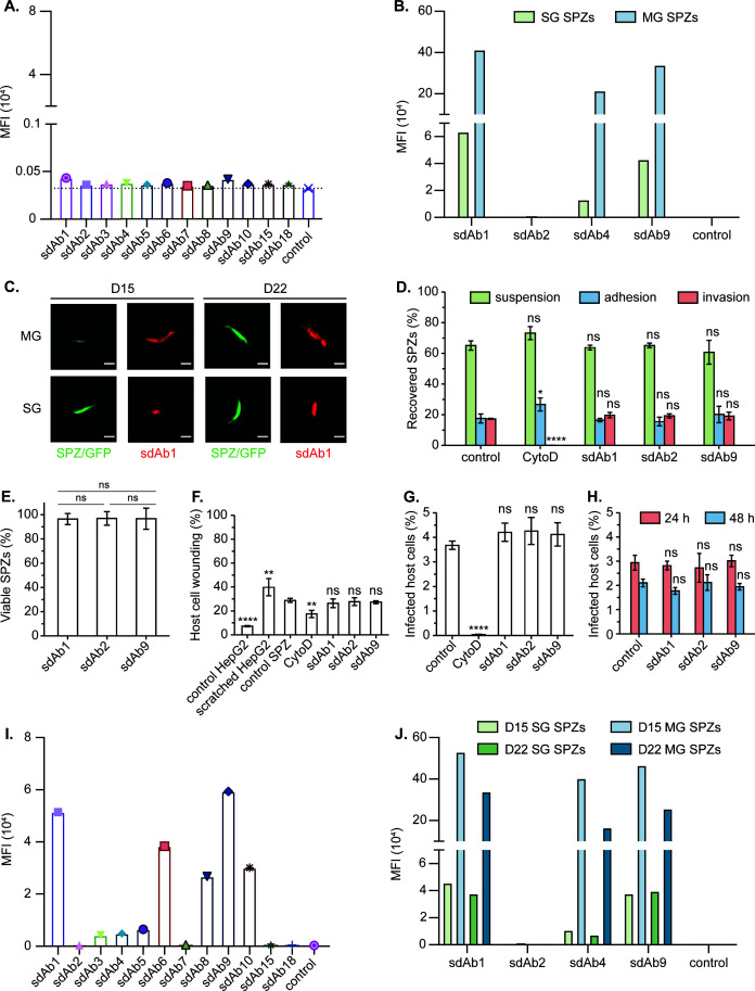
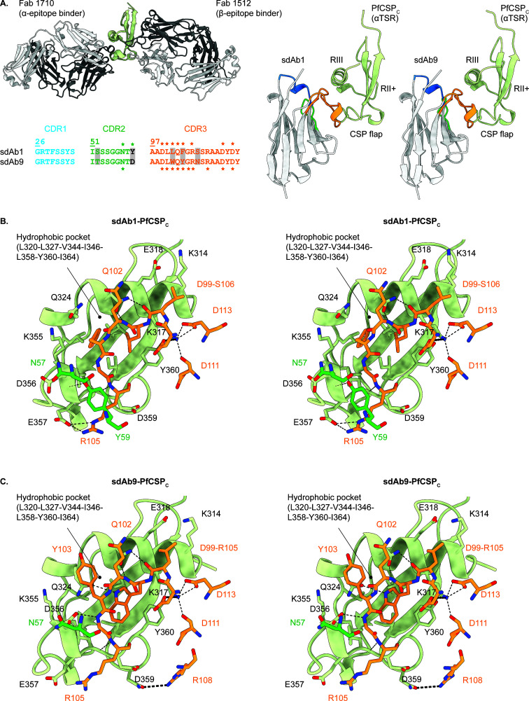
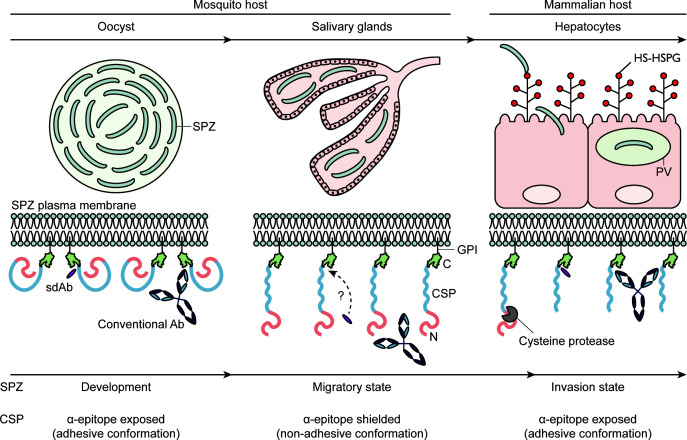
Plasmodium sporozoites (SPZs) are formed in the Anopheles mosquito midgut from where they travel to the salivary glands and subsequently to the mammalian liver after deposition into the skin. The SPZ's main surface antigen, the circumsporozoite protein (CSP), plays a pivotal role in SPZ biology and constitutes the immunodominant target for host antibodies. In this study, we raised single-domain antibodies (sdAbs) against CSP from P. falciparum (PfCSP) by immunizing two alpacas with recombinant versions of the antigen. We found that all identified sdAbs specifically target PfCSP's globular [Formula: see text]TSR domain without cross-reacting with P. berghei CSP. Further characterization revealed that most sdAbs recognize native PfCSP on the SPZ surface, although they do not have any inhibitory effect on hepatocyte binding and invasion. Structural studies showed that all binders target the previously identified [Formula: see text]-epitope, confirming the non-protective nature of this epitope. Comparison of sdAb binding to midgut and salivary gland SPZs revealed a shift in the exposure and accessibility of the [Formula: see text]-epitope. Hence, our findings provide further evidence that CSP undergoes structural changes during SPZ development in the mosquito host.

Keywords: Plasmodium falciparum; camelid single-domain antibodies; circumsporozoite protein; malaria; sporozoite.


Conflict of interest statement

The authors declare no conflict of interest.

Figures

Fig 1
Fig 1
Generation of PfCSP-specific sdAbs. (A) Schematic overview of the wild-type (WT) PfCSP from P. falciparum NF54 and the recombinant protein constructs derived from it shown in the left panel. The start of each domain and the segment corresponding to the globular αTSR domain are indicated on top and below the WT schematic, respectively. A legend of the conserved regions, tandem repeats, and purification tags (TEV, tobacco etch virus protease cleavage site; His6, hexahistidine tag) is shown at the bottom. An overview of the alpaca immunization strategy used to generate two immune VHH libraries is provided in the right panel. (B) Serum ELISA. Identification of HCAbs in the sera of Attitude (left panel) and Cosimo (right panel) targeting recombinant PfCSPFL and PfCSPC. (C) Cross-reactivity ELISA. The sdAbs raised against PfCSP were probed for binding to PfCSPFL and PbCSPFL (blue and orange bars, respectively). (B, C) The mean and standard deviation of triplicates for each condition are shown.
Fig 2
Fig 2
Heparin binding experiments with sdAb–PfCSPFL samples. The experimental setup is schematically illustrated in the top left panel. All collected fractions were analyzed by SDS-PAGE. Unbound PfCSPFL and sdAb were included as positive and negative controls, respectively.
Fig 3
Fig 3
In vitro interaction studies between the anti-PfCSP sdAbs and transgenic PfCSP–PbSPZs. (A) Interaction screening of the sdAbs to 20-day-old SG SPZs on ice. PBS was used as a negative control. (B) Interaction studies of selected sdAbs to 15-day-old MG and SG SPZs at ambient temperature. sdAb-BcII10 was included as a negative control. (C) Fluorescence imaging of MG and SG SPZs after incubation with sdAb1 at ambient temperature. Scale bars (white lines): 5 µm. (D–G) Effect of the sdAbs on SPZ invasion of HepG2 cells evaluated 2 h after parasite addition. Here, we measured the recovery of SPZs present in the suspension, attached to, or inside the cells (D), percentage of viable SPZs compared to the control (E), percentage of wounded cells (F), and percentage of infected cells (G). sdAb-BcII10 and CytoD were included as negative and positive controls for invasion inhibition, respectively. (H) Effect of the sdAbs on the SPZ intracellular development. The percentage of parasite-infected HepG2 cells 24 and 48 h post-incubation with SPZs pre-incubated with the different sdAbs is shown. sdAb-BcII10 was included as a negative control. (I) Interaction screening of the sdAbs to 13-day-old MG SPZs on ice. PBS was included as a negative control. (J) sdAb binding to MG and SG SPZs with different maturation times (15-day-old vs. 22-day-old) at ambient temperature. sdAb-BcII10 was included as a negative control. (A, B and I, J) Binding is expressed as median fluorescence intensities (MFI). (D–H) The mean values with standard deviation of three independent measurements are shown. Statistical significance was determined by one-way analysis of variance with Holm–Šídák correction for multiple comparisons; *P ≤ 0.05, **P ≤ 0.01, ****P ¡ 0.0001, ns = not significant.
Fig 4
Fig 4
Crystal structures of sdAb1 and sdAb9 in complex with PfCSPC. (A) Cartoon representations of PfCSPC (in light green) in complex with antibody fragments (in gray). The top left panel displays a superposition of the Fab1710-PfCSPC (PDB ID 6B0S [30]) and Fab1512-PfCSPC (PDB ID 7RXP [31]) crystal structures. The right panel shows the crystal structures of PfCSPC bound by sdAb1 and sdAb9 (PDB IDs 9HZJ and 9HZK, respectively, this work). A sequence alignment of the CDRs from sdAb1 and sdAb9 is presented in the bottom left panel, with amino acid substitutions highlighted by gray boxes. The CDRs are indicated in different colors (CDR1, blue; CDR2, green; CDR3, orange), and the residues that are part of the paratope are marked by an asterisk (*). Stereo views of the sdAb1–PfCSPC (B) and sdAb9–PfCSPC (C) interactions. PfCSPC is depicted in cartoon representation. For reasons of clarity, only the sdAb residues that are part of the paratope are shown and colored as in (A). All interacting residues are labeled and shown in stick representation. Hydrogen bonds and salt bridges are indicated by black dashed lines.
Fig 5
Fig 5
CSP is subject to conformational changes during SPZ development in the mosquito and its journey to the mammalian liver. CSP’s α-epitope is exposed during SPZ development in the mosquito midgut (adhesive conformation). Upon release from the oocyst, the SPZs transition to a migratory state and travel to the salivary glands. This transition is marked by a conformational change of CSP, rendering the α-epitope inaccessible to Abs and fragments thereof (non-adhesive form). sdAbs are expected to display better penetration properties due to their smaller size. Following deposition into the mammalian skin, the SPZs migrate to the liver, where they are primed for hepatocyte invasion through the interaction between CSP and highly sulfated heparan sulphate proteoglycans (HS-HSPGs). This interaction triggers the proteolytic cleavage of CSP by a cysteine protease, causing it to revert to its adhesive conformation and expose its α-epitope. Consequently, the SPZ switches to its invasion state, allowing it to invade a final hepatocyte and settle within a parasitophorous vacuole (PV) for its further development.

Similar articles

References

    1. WHO . 2024. World malaria report 2024: addressing inequity in the global malaria response
    1. Cowman AF, Healer J, Marapana D, Marsh K. 2016. Malaria: biology and disease. Cell 167:610–624. doi: 10.1016/j.cell.2016.07.055 - DOI - PubMed
    1. Kozlov M. 2021. Resistance to front-line malaria drugs confirmed in Africa. Nature New Biol 597:604–604. doi: 10.1038/d41586-021-02592-6 - DOI
    1. Normile D. 2021. Threat of drug-resistant malaria looms in Africa. Science 373:1427–1427. doi: 10.1126/science.acx9160 - DOI - PubMed
    1. Draper SJ, Sack BK, King CR, Nielsen CM, Rayner JC, Higgins MK, Long CA, Seder RA. 2018. Malaria vaccines: recent advances and new horizons. Cell Host Microbe 24:43–56. doi: 10.1016/j.chom.2018.06.008 - DOI - PMC - PubMed

Publication types

LinkOut - more resources