. 2025 May 6;122(18):e2501331122.
doi: 10.1073/pnas.2501331122. Epub 2025 Apr 28.

Dopamine induces fear extinction by activating the reward-responding amygdala neurons

Affiliations

Dopamine induces fear extinction by activating the reward-responding amygdala neurons

Xiangyu Zhang et al. Proc Natl Acad Sci U S A. .

Abstract

The extinction of conditioned fear responses is crucial for adaptive behavior, and its impairment is a hallmark of anxiety disorders such as posttraumatic stress disorder. Fear extinction takes place when animals form a new memory that suppresses the original fear memory. In the case of context-dependent fear memory, the new memory is formed within the reward-responding posterior subset of basolateral amygdala (BLA) that is genetically marked by Ppp1r1b+ neurons. These memory engram cells suppress the activity of the original fear-responding Rspo2+ engram cells present in the anterior BLA, hence fear extinction. However, the neurological nature of the teaching signal that instructs the formation of fear extinction memory in the Ppp1r1b+ neurons is unknown. Here, we demonstrate that ventral tegmental area (VTA) dopaminergic signaling drives fear extinction in distinct BLA neuronal populations. We show that BLA fear and extinction neuronal populations receive topographically divergent inputs from VTA dopaminergic neurons via differentially expressed dopamine receptors. Fiber photometry recordings of dopaminergic activity in the BLA reveal that dopamine (DA) activity is time-locked to freezing cessation in BLA fear extinction neurons, but not BLA fear neurons. Furthermore, this dopaminergic activity in BLA fear extinction neurons correlates with extinction learning. Finally, using projection-specific optogenetic manipulation, we find that activation of the VTA DA projections to BLA reward and fear neurons accelerated or impaired fear extinction, respectively. Together, this work demonstrates that dopaminergic activity bidirectionally controls fear extinction by distinct patterns of activity at BLA fear and extinction neurons.

Keywords: amygdala; dopamine; fear extinction; reward; valence.


Conflict of interest statement

Competing interests statement:The authors declare no competing interest.

Figures

Fig. 1.
Fig. 1.
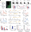
Anterograde and retrograde tracing of VTA dopaminergic neurons to BLA. (A) Schematic of AAV9-DIO-ChR2-EYFP injection into VTA of DAT-IRES-Cre mice. (B) Left, representative image of ChR2-EYFP expression in the VTA, TH in red. Right, confocal images of anterograde EYFP-expressing fibers (green, Left Top and Bottom) through the medial–lateral axis of the BLA (red, Middle-Top Ppp1rb1 and Middle-Bottom Ppp1rb1+ subregions; overlap on Right Top-Bottom panels, blue DAPI). (C) Quantification of mean pixel intensity in the LA, aBLA, and pBLA in arbitrary units (N = 3 replicate mice, n = 18 sections per region; One-way ANOVA, ***P < 0.001). (D) Schematic of helper and rabies virus injection into aBLA of Rspo2-Cre mice. (E) Representative confocal images of rabies-mediated retrograde tracing from BLA Rspo2+ neurons to VTA dopaminergic neurons (Inset: I = TH+ and II = TH neurons projecting to Rspo2+ neurons). (F) Schematic of helper and rabies virus injection into pBLA of Ppp1rb1-Cre mice. (G) Representative confocal images of rabies-mediated retrograde tracing from pBLA Ppp1r1b+ neurons to VTA dopaminergic neurons (Inset: I = TH+ and II = TH neurons projecting to Ppp1r1b+ neurons). (H) Bilateral rostro-caudal distribution of VTA dopaminergic cells projecting to Rspo2+ (green, n = 208 cells) or Ppp1r1b+ (red, n = 101 cells) neurons (χ2 < 0.0001, Rspo2-Cre mice N = 4, Ppp1r1b-Cre mice: N = 3, injections in the right hemisphere).
Fig. 2.
Fig. 2.
Quantification of D1 and D2 dopamine receptor expression in the BLA. (A) Double smFISH of Drd1 (green) and Drd2 (red) in BLA along the AP axis. (B) Fourplex smFISH of Drd1 (white) and Drd2 (yellow), Rspo2 (green, and green dotted line) and Ppp1rb1 (red, and red dotted line) in BLA. (C) Left: high magnification images showing colocalization of neurons containing both Drd1 and Rspo2 or Ppp1r1b mRNA in BLA. Right: normalized count of double positive cells (Rspo2-Drd1 green, and Ppp1r1b-Drd1 red) plotted as a function of the Drd1 mRNA fluorescence intensity. Rspo2 cells: n = 272; Ppp1r1b cells: n = 325. Unpaired t test, P = 2.5*10^6.
Fig. 3.
Fig. 3.
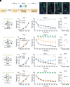
Dopamine activity in BLA Ppp1r1b+ neurons is correlated with extinction learning. (A) Schematic of the fiber photometry implant. (B) Confocal image displaying aBLA of Rspo2-Cre mouse expressing the dopamine sensor GRABda (green) and fiber optic track (squared box). (C) Schematic of the experimental setup: 35 d after surgery, mice were habituated to the photometry setup and connected to the patch cord for 20 min in the home cage, and underwent an open field test, contextual fear conditioning, extinction, and extinction recall. (D) Group average freezing levels after shock (Left), during extinction (Middle), and extinction recall during first 3 min (Right). (E) Group average dopamine activity to shock onset for three shock trials for Rspo2+ neurons. (F) Example traces of photometry signals (reported as z-score) during early (Top) and late (Bottom) extinction in Rspo2+ neurons. Yellow boxes above the traces indicate freezing bouts (0.5 s > light yellow < 2 s; dark yellow >= 2 s). (G) Dopamine activity aligned to transition from freezing (>0.5 s) to movement (>3 s) during early and late extinction in Rspo2+ neurons. (H) Group average dopamine activity to shock onset for three shock trials for Ppp1r1b+ neurons. (I) Example traces of photometry signals (reported as z-score) during early (Top) and late (Bottom) extinction in Ppp1r1b-Cre mice. Yellow boxes above the traces indicate freezing bouts. (J) Dopamine activity aligned to transition from freezing (>0.5 s) to movement (>3 s) during early and late extinction in Ppp1r1b+ neurons. (K) Postpeak area under the curve for each shock. Rspo2+ neurons display a response stronger than Ppp1r1b+ neurons (unpaired t test, **P < 0.01). (L) Quantification of the peak z-score observed in the 3 s after freezing cessation in Rspo2+ andPpp1r1b+ neurons in early and late extinction (unpaired t test, **P < 0.01, ***P < 0.001). Ppp1r1b+ neurons display a response stronger than Rspo2+ neurons. (M) Left: correlation between the average peak z-score observed in the 3 s after freezing cessation in Rspo2+ and Ppp1r1b+ neurons during early extinction and the change in percent freezing from early to late extinction (Spearman’s correlation: Rspo2, R = 0.31, P = 0.46; Ppp1r1b, R = 0.88, P = 0.0039). Right: correlation between the average peak z-score observed in the 3 s after freezing cessation in Rspo2+ and Ppp1r1b+ neurons during late extinction and the change in percent freezing from early to late extinction (Spearman’s correlation: Rspo2, R = 0.11, P = 0.82; Ppp1r1b, R = −0.24, P = 0.57). For all panels: n = 8 Rspo2-Cre, n = 8, Ppp1r1b-Cre. **P < 0.01; ***P < 0.001. Error bars or shaded regions indicate SEM.
Fig. 4.
Fig. 4.
Bidirectional control of fear extinction by optogenetic manipulation of VTA Dopaminergic projection to pBLA or aBLA. (A) Experimental protocol of optogenetic manipulation during fear extinction training. (B) Confocal images showing fiber optic placement and ChR2-YFP+ or NpHR3.0-EYFP+ DA axons in anterior and pBLA in relation to schematics (C, E, G, and I), respectively. (C) Schematic of AAV-DIO-ChR2-EYFP injection into VTA and optical fiber implant targeting pBLA of DAT-IRES-Cre mice. (D) Optogenetic activation of VTA dopaminergic projections to pBLA facilitated fear extinction learning and fear extinction memory. EYFP group n = 9; ChR2 group n = 10. Day1 and Day 2: Two-way RM ANOVA; Day 3: Unpaired t test. (E) Schematic of AAV-DIO-NpHR3.0-EYFP injection into VTA and optical fiber implant targeting pBLA of DAT-IRES-Cre mice. (F) Optogenetic inhibition of VTA dopaminergic projections to pBLA impaired fear extinction learning and fear extinction memory. EYFP group n = 11; NpHR group n = 10. Day 1 and Day 2: Two-way RM ANOVA; Day 3: Unpaired t test. (G) Schematic of AAV-DIO-ChR2-EYFP injection into VTA and optical fiber implant targeting aBLA of DAT-IRES-Cre mice. (H) Optogenetic activation of VTA dopaminergic projections to aBLA suppressed fear extinction learning and fear extinction memory. EYFP group n = 8; ChR2 group n = 8. Day 1 and Day 2: Two-way RM ANOVA; Day 3: Unpaired t test. (I) Schematic of AAV-DIO-NpHR3.0-EYFP injection into VTA and optical fiber implant targeting aBLA of DAT-IRES-Cre mice. (J) Optogenetic inhibition of VTA dopaminergic projections to aBLA did not affect fear extinction behavior. EYFP group n = 9; NpHR group n = 8. Day 1 and Day 2: Two-way RM ANOVA; Day 3: Unpaired t test. Data are presented as mean ± SEM. *P < 0.05, **P <0.01, ***P <0.001, ****P <0.0001.
Fig. 5.
Fig. 5.
Manipulation of Drd1 in Rspo2 and Ppp1r1b neurons. (A) Schematic of the experimental procedure. An AAV-Ef1a-DIO-Drd1-EYFP or an AAV5-CAG-DIO-mDrd1a-shRNAmir injected in the pBLA or aBLA of Rspo2 pr Ppp1r1b Cre mice to induce Drd1 overexpression or KD respectively. (B) Conditioning protocol. (C) Drd1 mRNA expression in pBLA for control (Ctrl) and overexpressing (OE) conditions. Unpaired t test, ***P < 0.001, **P < 0.01. (D) Confocal images showing D1 receptor overexpression in pBLA. (E) Drd1 mRNA expression in pBLA for Ctrl and KD conditions. Unpaired t test, ***P < 0.001, **P < 0.01. (F) Confocal images showing D1 receptor KD in pBLA. (G) Drd1 overexpression (OE) in BLA Ppp1r1b+ neurons caused significantly lower freezing level on Day 2 and Day 3. (H) Drd1 KD impaired extinction learning (Day 2) and memory (Day 3). (I) Drd1 overexpression (OE) in BLA Rspo2+ neurons did not cause a change in freezing level on Day 2 and Day 3. (J) Drd1 KD in BLA Rspo2+ did not impaired extinction learning (Day 2) and memory (Day 3). Two-way ANOVA, *P < 0.05, **P < 0.01, ***P < 0.001, ****P < 0.0001. Data are presented as mean ± SEM.
Fig. 6.
Fig. 6.
Role of VTA DA innervation to Rspo+ and Ppp1r1b+ BLA neurons during fear conditioning and fear extinction. (A) Schematic model of the circuit activity in relation to fear conditioning and fear extinction. During fear conditioning (Left, Top to Bottom), the US (electric shock, red) causes DA release on both Rspo2+ (red) and Ppp1r1b+ (blue) BLA neurons engaged in a competitive inhibitory antagonism. The DA release is larger on Rspo2+ BLA neurons, which in turn inhibit Ppp1r1b+ BLA neurons promoting freezing. During fear extinction (Right, Top to Bottom), the lack of US causes DA release mainly on Ppp1r1b+ BLA neurons, which in turn inhibit Rspo2+ BLA neurons promoting freezing cessation.

References

    1. Shalev A., Liberzon I., Marmar C., Post-traumatic stress disorder. N. Engl. J. Med. 376, 2459–2469 (2017). - PubMed
    1. Martin E. I., Ressler K. J., Binder E., Nemeroff C. B., The neurobiology of anxiety disorders: Brain imaging, genetics, and psychoneuroendocrinology. Psychiatr. Clin. North Am. 32, 549–575 (2009). - PMC - PubMed
    1. Pavlov I. P., Conditioned Reflexes (Oxford University Press, 1927).
    1. Zhang X., Kim J., Tonegawa S., Amygdala reward neurons form and store fear extinction memory. Neuron 105, 1077–1093.e7 (2020). - PubMed
    1. Grewe B. F., et al. , Neural ensemble dynamics underlying a long-term associative memory. Nature 543, 670–675 (2017). - PMC - PubMed

LinkOut - more resources