. 2025 May 6;122(18):e2421391122.
doi: 10.1073/pnas.2421391122. Epub 2025 Apr 28.

Water-directed pinning is key to tau prion formation

Affiliations

Water-directed pinning is key to tau prion formation

Michael P Vigers et al. Proc Natl Acad Sci U S A. .

Abstract

Tau forms fibrillar aggregates that are pathological hallmarks of a family of neurodegenerative diseases known as tauopathies. The synthetic replication of disease-specific fibril structures is a critical gap for developing diagnostic and therapeutic tools. This study debuts a strategy of identifying a critical and minimal folding motif in fibrils characteristic of tauopathies and generating seeding-competent fibrils from the isolated tau peptides. The 19-residue jR2R3 peptide (295 to 313) which spans the R2/R3 splice junction of tau, and includes the P301L mutation, is one such peptide that forms prion-competent fibrils. This tau fragment contains the hydrophobic VQIVYK hexapeptide that is part of the core of all known pathological tau fibril structures and an intramolecular counterstrand that stabilizes the strand-loop-strand (SLS) motif observed in 4R tauopathy fibrils. This study shows that P301L exhibits a duality of effects: it lowers the barrier for the peptide to adopt aggregation-prone conformations and enhances the local structuring of water around the mutation site to facilitate site-directed pinning and dewetting around sites 300-301 to achieve in-register stacking of tau to cross β-sheets. We solved a 3 Å cryo-EM structure of jR2R3-P301L fibrils in which each protofilament layer contains two jR2R3-P301L copies, of which one adopts a SLS fold found in 4R tauopathies and the other wraps around the SLS fold to stabilize it, reminiscent of the three- and fourfold structures observed in 4R tauopathies. These jR2R3-P301L fibrils are competent to template full-length 4R tau in a prion-like manner.

Keywords: DEER; cryo-EM; neurodegenerative disease; protein aggregation; tauopathy.


Conflict of interest statement

Competing interests statement:K.S.K. consults for ADRx and Expansion Therapeutics and is a member of the Tau Consortium Board of Directors, M.P.V., S.L., A.D., A.P.L., K.T., K.S.K., and S.H. have filed for a patent based on the design of tau peptides presented in this paper. Patent information: Disc-ID-24-06-12-001.

Figures
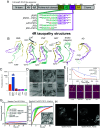
Fig. 1.
Fig. 1.
Fibril formation and stability of jR2R3 peptides: (A) (Upper) The domains of the longest isoform of tau, consisting of an N-terminal domain, the N1 and N2 repeat domains, a proline-rich domain, the four repeat domains R1-R4 and a C-terminal domain. (Lower) Sequence of the jR2R3 peptide variants. (B) Tauopathy fibril structures: CBD type II (PDB#: 6TJX), LNT (PDB #: 7P6a), PSP (PDB #: 7P65), GGT type I (PDB #: 7P66), 0N4R Snake (PDB #: 6QJH), and AD PHF (PDB #: 5O3L). (C) Maximum ThT fluorescence after incubation with heparin at 37 °C for 18 h. Samples contained 50 µM protein with 12.5 µM heparin (n = 3). (D) nsTEM of all jR2R3 variant fibrils formed with heparin. (Scale bar, 100 nm.) (E) ThT fluorescence of fibrils after incubation with guanidinium hydrochloride (GdnHCl) at 37 °C for 18 h (n = 3). (F) AFM images of denatured samples. Fibrils degrade in a GdnHCl concentration dependent manner. (G) Seeding of tau187-P301L with jR2R3 fibrils (n = 3). Left, raw ThT fluorescence. Right, normalized ThT to highlight kinetics of aggregation of the aggregating reactions. Inset: nsTEM of fibrils seeded with jR2R3-P301L fibrils. (Scale bar, 1 µm.) (H) Transfection of H4 cells expressing tau187 with jR2R3-(P301L). Endogenous tau accumulates in aggregates upon transfection with jR2R3-P301L, but not jR2R3.
Fig. 2.
Fig. 2.
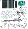
Structure of jR2R3-P301L fibrils: (A) Example cryo-EM image of jR2R3-P301L fibrils. (B) Representative 2D class average of the singlet class of fibrils. (Left) initial class used for initial model building. (Right) 2D projection after refinement. (C) EM map of the fibril viewed from the side and down the axis of the fibril colored by the estimated resolution of the map. (D) Atomic structure of a single jR2R3-P301L fibril layer (PDB ID #8V1N). (E) Schematic of the jR2R3-P301L fibril layer. Pink dots are glycine residues, blue dots are positively charged side chains, red are negatively charged, green are hydrophilic, and white are hydrophobic side chains. (F) Structure of the jR2R3-P301L inner strand–loop–strand (SLS) chain alongside other conformations of the segment in tauopathy structures. H299 and Y310 are shown in yellow to highlight differences in the orientation of side chains across the structures.
Fig. 3.
Fig. 3.
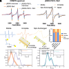
Electron paramagnetic resonance of jR2R3 and jR2R3-P301L. (A) CW EPR spectra of jR2R3 and jR2R3-P301L before and after fibrillization. (B) Simulated spectra of the 3 components used to fit (A). (C) Examples of the types of species that contribute to the three components used to fit CW EPR spectra. (D) The proportion of the spectra attributed to mobile (blue), immobile (orange), and immobile, spin-exchanging, species (yellow). (E) DEER probability distribution [P(r)] of jR2R3 (294 to 305) and jR2R3-P301L (294 to 304) fibrils. The expected P(r) of jR2R3 in the LNT and CBD conformation is shown in dashed black and red respectively. Time domain signal is shown in inset. (F) DEER P(r) comparing the jR2R3-P301L fibril formed with heparin (purple), to the jR2R3-P301L fibrils formed by seeding with jR2R3-P301L heparin fibrils (teal).
Fig. 4.
Fig. 4.
(A) Free energy landscapes of jR2R3 and jR2R3-P301L from α-carbon distances in REMD simulations show differences in energy well depths and pathways to opening. (i–vi) Six jR2R3-P301L clusters from the boxed regions of the energy landscape; balls denote K298, and Q307 (black) and V300 and S305 (red). Figure 4A: Modified with permission from ref. , which is licensed under CC BY-NC-ND. (B) jR2R3-P301L forms more oligomers with 5 or more intermolecular backbone hydrogen bonds in dimer simulations than jR2R3, but these oligomer conformations rarely occur in dimers where one of the monomers is clamped and pinched. Error bars show 90% CI. (C) The backbone flexibility quantified by the residue backbone entropy of each residue in jR2R3 and jR2R3-P301L (details in Supplement). A decrease in the entropy from residues V300 and P301L is seen in jR2R3. (D) jR2R3-P301L has more tetrahedral water molecules than jR2R3 around residue 301; this tetrahedral water fraction is associated with slower water and more hydrophobicity. (E) kσ of jR2R3 and jR2R3-P301L from the N terminus (294C), V300C, and at the C-terminus 314C of each peptide. A significant reduction in dynamics was observed at the V300C of jR2R3-P301L in comparison the V300C of jR2R3 and 294C of jR2R3-P301L. All other comparisons were insignificant (P > 0.05) with an independent t test. n ≥ 3 for all samples. (F) The dewetting free energy per water molecule of P301 and 301L residues, Nw and N0, are the instantaneous and equilibrium number of water molecules in the probe volume, respectively. (G) Representation of the dewetting process that is driven by the release of hydration water.

References

    1. Jensen M. R., et al. , Quantitative determination of the conformational properties of partially folded and intrinsically disordered proteins using NMR dipolar couplings. Structure 17, 1169–1185 (2009). - PubMed
    1. Mandelkow E. M., Mandelkow E., Biochemistry and cell biology of Tau protein in neurofibrillary degeneration. Cold Spring Harb. Perspect. Biol. 2, a006247 (2011). - PMC - PubMed
    1. Wang Y., Mandelkow E., Tau in physiology and pathology. Nat. Rev. Neurosci. 17, 5–21 (2016). - PubMed
    1. Fitzpatrick A. W. P., et al. , Cryo-EM structures of tau filaments from Alzheimer’s disease. Nature 547, 185–190 (2017). - PMC - PubMed
    1. Arakhamia T., et al. , Posttranslational modifications mediate the structural diversity of tauopathy strains. Cell 180, 633–644.e12 (2020). - PMC - PubMed

LinkOut - more resources