. 2025 Apr 28;9(17):CASE24850.
doi: 10.3171/CASE24850. Print 2025 Apr 28.

Improbable discovery of an incidental high-grade AVM: illustrative case

Affiliations

Improbable discovery of an incidental high-grade AVM: illustrative case

Max S Fleisher et al. J Neurosurg Case Lessons. .

Abstract

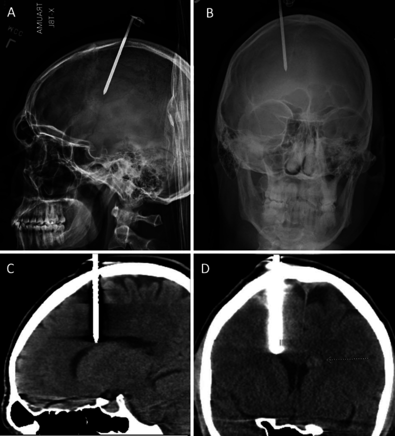
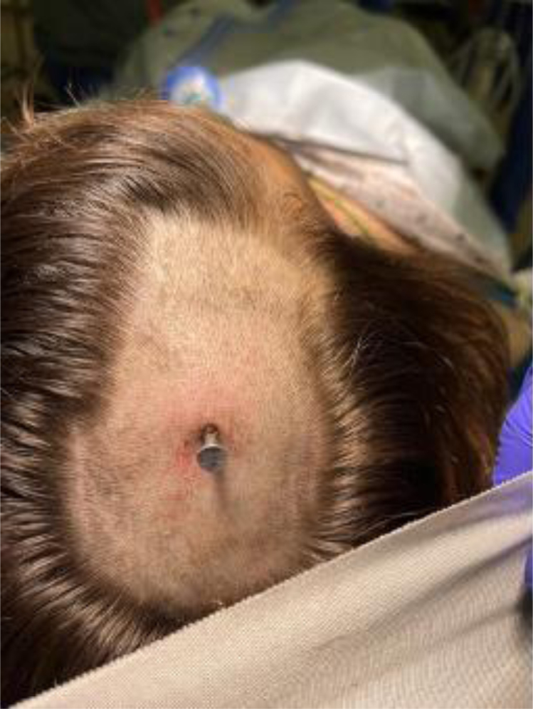
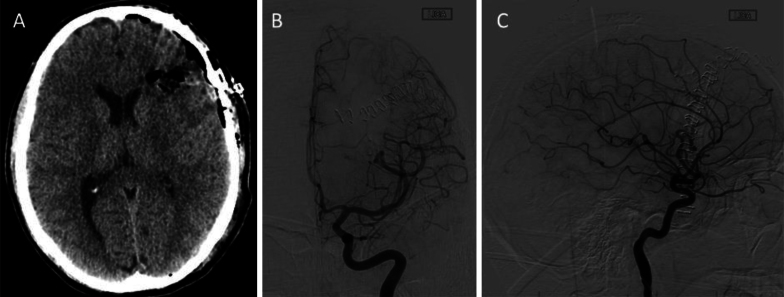
Background: The authors discuss the first reported case of a large, high-grade arteriovenous malformation (AVM) in the dominant hemisphere, discovered incidentally after a penetrating nail gun injury.

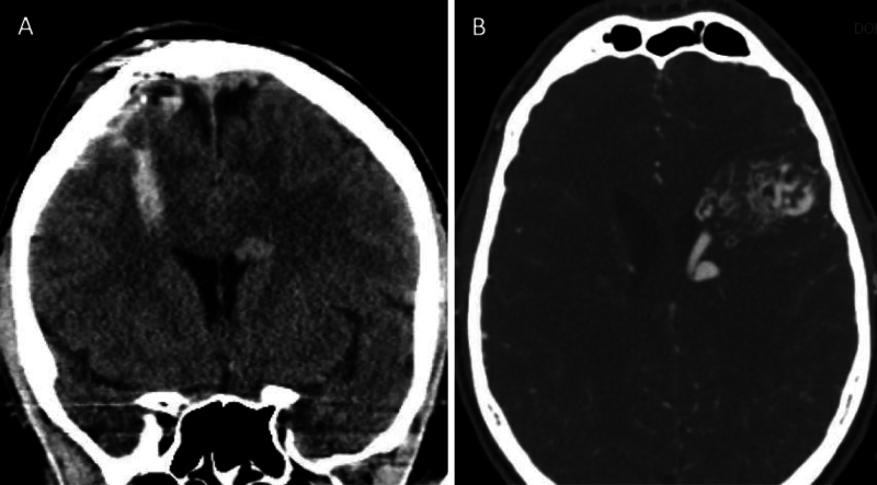
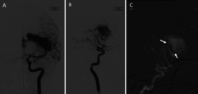
Observations: The patient underwent surgical removal of a nail lodged in the right frontal lobe. A contralateral AVM was diagnosed on his perioperative imaging and was evaluated further with diagnostic cerebral angiography. Because of the location of the AVM within the dominant fronto-opercular region, the patient underwent a super-selective Wada test to evaluate for the risk of expressive language deficit prior to undergoing a successful resection of the AVM. He had an excellent recovery from both surgeries without any neurological deficits.

Lessons: This case illustrates the importance of continued suspicion for incidental findings when reviewing imaging, despite the presence of a known and obvious pathology. The observations add nuance to the standard considerations for surgical intervention for penetrating nail gun injuries, and the workup for incidentally found vascular lesions is reviewed. https://thejns.org/doi/10.3171/CASE24850.

Keywords: cerebral arteriovenous malformation; incidental finding; nail gun injury; penetrating traumatic brain injury.


Figures

FIG. 1.
FIG. 1.
Preoperative imaging. Lateral (A) and AP (B) radiographs showing the hyperdense foreign object from the nail gun injury. Sagittal (C) and coronal (D) CT images showing the nail trajectory near the right lateral ventricle. A small hyperdensity can be noted near the left lateral ventricle, hinting at an underlying pathology.
FIG. 2.
FIG. 2.
Photograph showing the nail gun injury site in the operating room, after hair was shaved at the site.
FIG. 3.
FIG. 3.
Postoperative imaging after removal of the nail. A: Coronal CT image without contrast showing a small amount of hemorrhage within the penetrating nail tract, as well as the suspicious left periventricular hyperdensity. B: Axial CT angiogram showing the large AVM within the left frontal lobe, with the 3.7-cm nidus centered near the pars opercularis.
FIG. 4.
FIG. 4.
Digital subtraction angiograms. AP (A) and lateral (B) injections of the left internal carotid artery showing a large left-sided AVM (Spetzler-Martin grade 4, supplemented grade 6) with feeders primarily from the left MCA. There is deep venous drainage into the left internal cerebral vein and vein of Galen. There is also superficial cortical drainage into the vein of Labbé. Lateral view (C) of a super-selective injection via the two left MCA pedicles (arrows), which were used for injection of methohexital and lidocaine for Wada provocative testing.
FIG. 5.
FIG. 5.
Postoperative imaging after resection of the AVM. Axial CT head scan without contrast (A) showing resection of the AVM without complication. AP (B) and lateral (C) digital subtraction angiograms showing complete resection of the AVM without signs of nidus filling or early venous drainage.

Similar articles

References

    1. Hafez A, Koroknay-Pál P, Oulasvirta E.The application of the novel grading scale (Lawton-Young grading system) to predict the outcome of brain arteriovenous malformation. Neurosurgery. 2019;84(2):529-536. - PMC - PubMed
    1. Can A Gross BA Du R.. The natural history of cerebral arteriovenous malformations. Handb Clin Neurol. 2017;143:15-24. - PubMed
    1. Bican O, Cho C, Lee L.Positive pharmacologic provocative testing with methohexital during cerebral arteriovenous malformation embolization. Clin Imaging. 2018;51:155-159. - PubMed
    1. Tawk RG Tummala RP Memon MZ Siddiqui AH Hopkins LN Levy EI.. Utility of pharmacologic provocative neurological testing before embolization of occipital lobe arteriovenous malformations. World Neurosurg. 2011;76(3-4):276-281. - PubMed
    1. Feliciano CE de León-Berra R Hernández-Gaitán MS Torres HM Creagh O Rodríguez-Mercado R.. Provocative test with propofol: experience in patients with cerebral arteriovenous malformations who underwent neuroendovascular procedures. AJNR Am J Neuroradiol. 2010;31(3):470-475. - PMC - PubMed

LinkOut - more resources