. 2025 May-Jun;39(3):1470-1491.
doi: 10.21873/invivo.13948.

First Experiences of Pilot Clinical Studies on Boron Neutron Capture Therapy for Recurrent Gastrointestinal Cancers Using an Intravenous Injection of 10BPA

Affiliations

First Experiences of Pilot Clinical Studies on Boron Neutron Capture Therapy for Recurrent Gastrointestinal Cancers Using an Intravenous Injection of 10BPA

Hironobu Yanagie et al. In Vivo. 2025 May-Jun.

Abstract

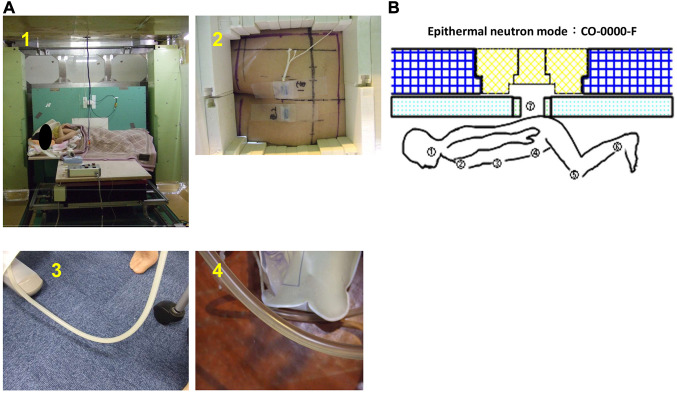
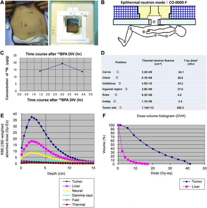
Background/aim: Boron neutron capture therapy (BNCT) is a novel treatment that induces targeted tumor cell damage through the selective accumulation of 10B compounds in cancer cells followed by the production of alpha and lithium particles using thermal neutron irradiation. Despite its potential, clinical applications of BNCT for recurrent gastrointestinal cancers remain limited. This study presents the first pilot clinical evaluation of BNCT using intravenous boronophenylalanine (10BPA) for such cancers.

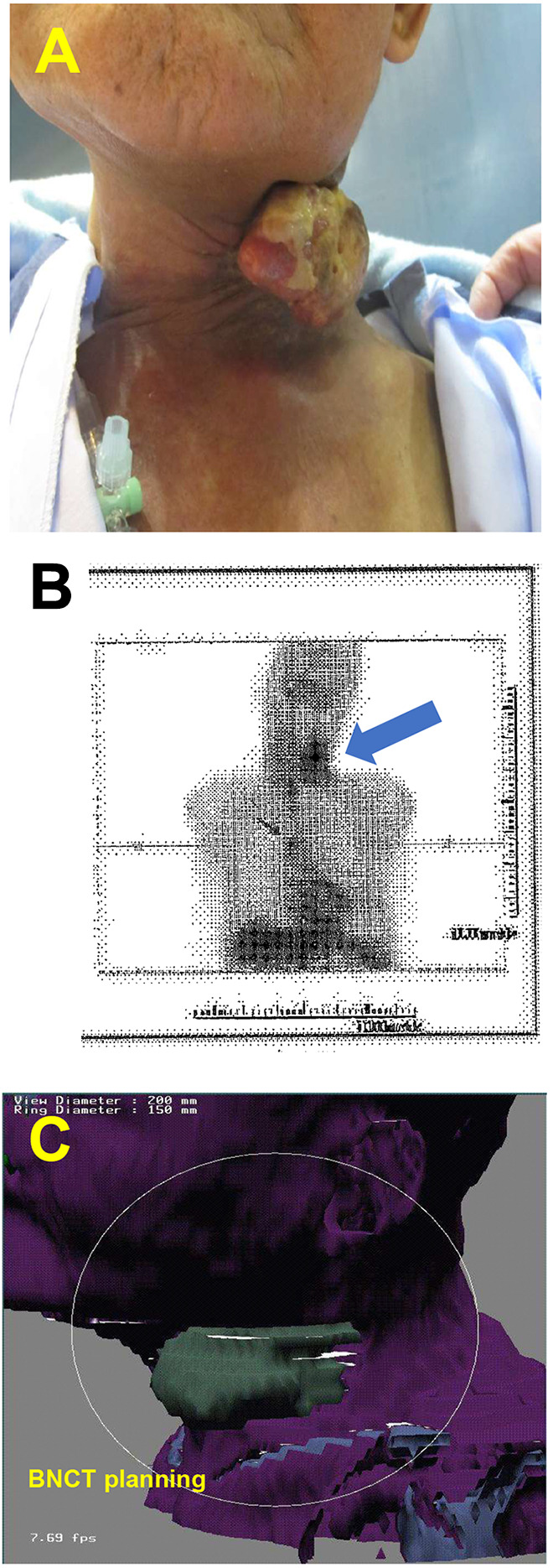
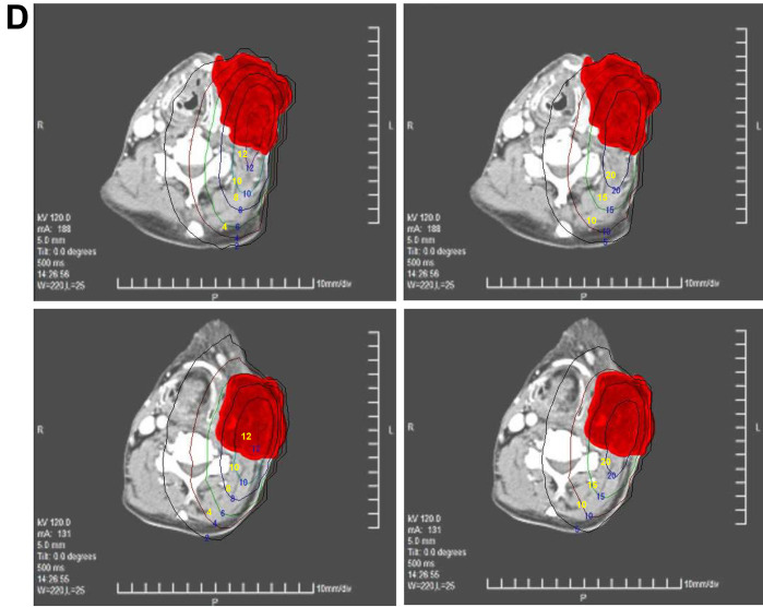
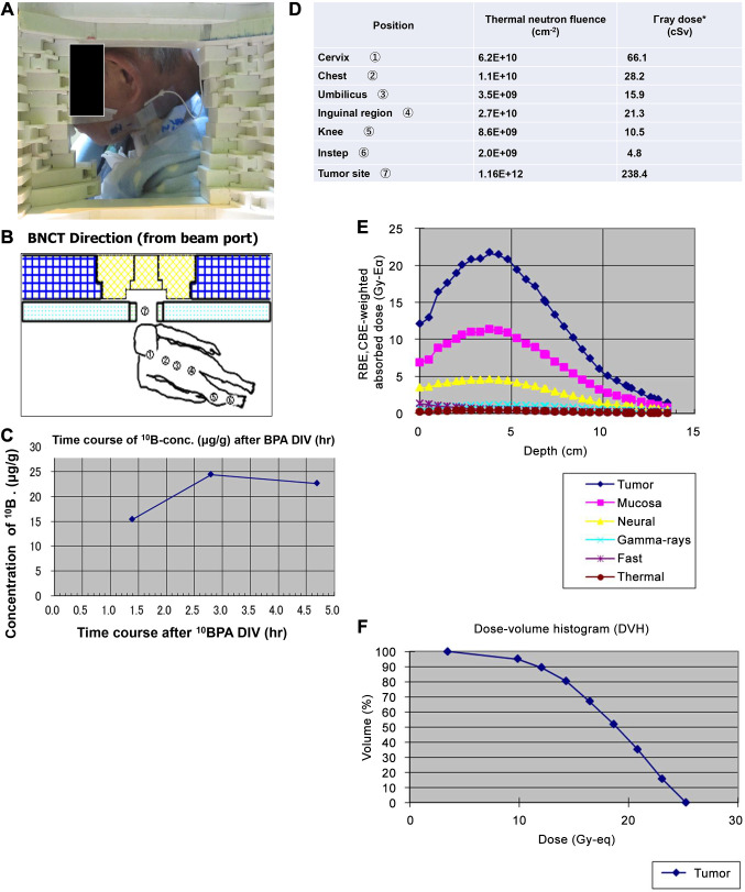
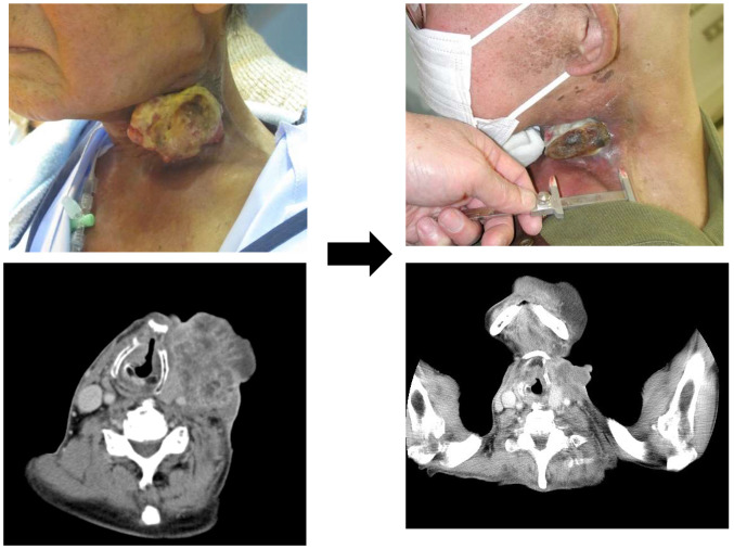
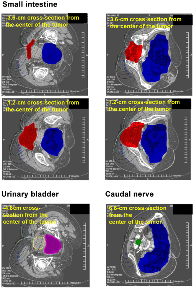
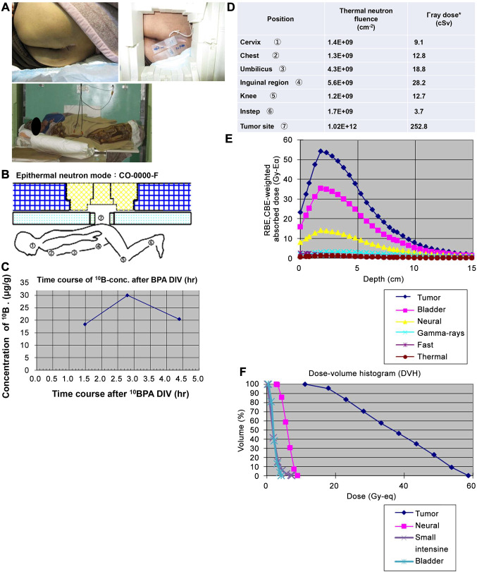
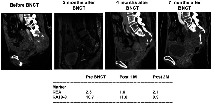
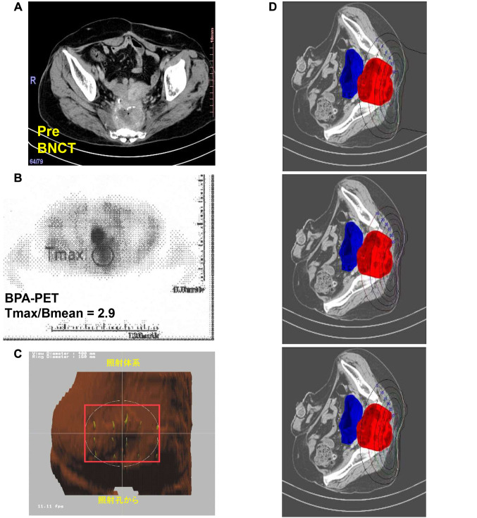
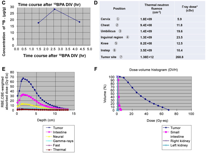
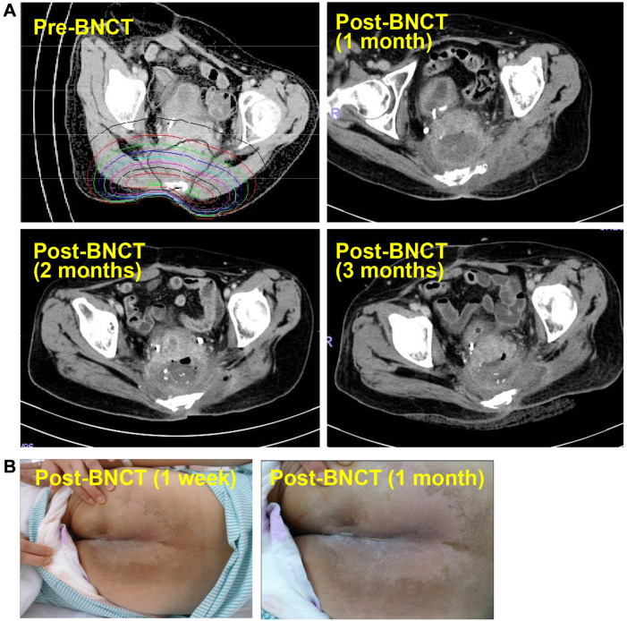
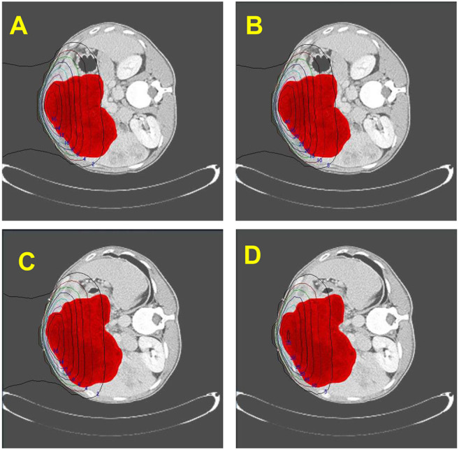
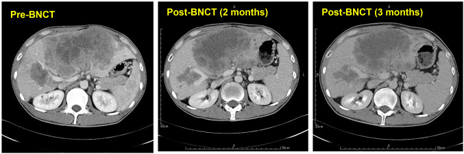
Case reports: Four patients with recurrent gastrointestinal cancers were enrolled in this phase I-II clinical study. All had tumors refractory to standard treatments, including surgery, chemotherapy, and radiotherapy. BNCT was performed using thermal neutron irradiation at Kyoto University Research Reactor. 10BPA was administered intravenously at 400 mg/kg, and no severe adverse effects were observed. Tumor responses varied, with one patient achieving partial response and three demonstrating stable disease at three months post-treatment. Notably, BNCT alleviated cancer-related symptoms, such as pain and nerve compression, improving patients' quality of life. Dosimetric evaluations confirmed effective tumor doses with acceptable exposure to surrounding normal tissues.

Conclusion: BNCT is a promising modality for recurrent gastrointestinal cancers, offering symptom relief and potential antitumor effects. Its safety and feasibility were demonstrated in this study. Future research should explore fractionated BNCT and combination therapies with immunotherapy or targeted agents to enhance efficacy further.

Keywords: Alpha particle; Boron neutron capture therapy (BNCT); Boronophenylalanine (10BPA); atomic reactor; recurrent gastrointestinal cancers; thermal neutron.


Conflict of interest statement

The Authors have no conflicts of interest to declare in relation to this study.

Figures

Figure 1
Figure 1
Imaging and treatment planning for boron neutron capture therapy (BNCT) in Case 1. (A) Photograph of the metastatic cervical gastric tumor before BNCT. (B) 18F-10BPA-PET image showing boron accumulation in the tumor. (C) Three-dimensional BNCT treatment plan visualized from the neutron beam port. (D) Two-dimensional distribution of thermal neutron flux (Gy-Eq/h) in the left cervical tumor following frontal epithermal neutron beam irradiation. The upper panel shows the -1.5 cm cross-section from the tumor center (left: dose to normal skin; right: dose to tumor; red: tumor tissue). The lower panel shows the 0.0 cm cross-section from the tumor center (left: dose to normal skin; right: dose to tumor; red: tumor tissue).
Figure 1
Figure 1
Imaging and treatment planning for boron neutron capture therapy (BNCT) in Case 1. (A) Photograph of the metastatic cervical gastric tumor before BNCT. (B) 18F-10BPA-PET image showing boron accumulation in the tumor. (C) Three-dimensional BNCT treatment plan visualized from the neutron beam port. (D) Two-dimensional distribution of thermal neutron flux (Gy-Eq/h) in the left cervical tumor following frontal epithermal neutron beam irradiation. The upper panel shows the -1.5 cm cross-section from the tumor center (left: dose to normal skin; right: dose to tumor; red: tumor tissue). The lower panel shows the 0.0 cm cross-section from the tumor center (left: dose to normal skin; right: dose to tumor; red: tumor tissue).
Figure 2
Figure 2
Patient positioning, neutron dosimetry, and dose distribution for boron neutron capture therapy (BNCT) in Case 1. (A) Photograph showing the body position of Case 1 for BNCT from the beam port. (B) Schematic representation of the body position for BNCT from the beam port. (C) Time course of changes in 10B concentration (μg/g) following intravenous infusion of 10BPA (absolute values in Case 1). (D) Measured thermal neutron fluence and gamma-ray dose delivered to the body. The measurable lower limits were approximately 5×107 cm−2 for thermal neutron fluence and ~0.01 cSv for gamma-ray dose (absolute values in Case 1). (E) Dose distribution along the beam axis in thermal neutron irradiation mode (CO-0000-F+ collimator, irradiation field: 15 cm×15 cm). Tumor: Tumor dose; Mucosa: oral mucosal dose; Neural: normal nerve dose; Gamma-rays: Gamma ray dose; Fast: fast neutron dose; Thermal: thermal neutron dose. (F) Calculated dose-volume histogram (DVH) of the tumor (tumor volume: 151.76 cm3). To achieve a tumor dose of 16 Gy at a depth of 5 cm, the BNCT irradiation time was set to 90 minutes.
Figure 3
Figure 3
Tumor response before and after boron neutron capture therapy (BNCT) in Case 1. Photographs and computed tomography (CT) images taken before (Left) and two months after BNCT (Right). Tumor growth was suppressed, and recanalization of the left internal jugular vein was observed following BNCT.
Figure 4
Figure 4
Two-dimensional distribution of neutron flux in the pelvic cavity in Case 2. Two-dimensional distribution of neutron flux (Gy-Eq/h) in the pelvic cavity. The upper and middle panels show the distribution at -3.6 cm, 3.6 cm, -1.2 cm, and 1.2 cm cross-sections from the tumor center (Gy-Eq), with red indicating the tumor and blue indicating the small intestine. The lower left panel shows the distribution at a -4.8 cm cross-section, with purple indicating the urinary bladder. The lower right panel shows the distribution at a 6.6 cm cross-section, with green indicating the caudal nerve and blue indicating the small intestine.
Figure 5
Figure 5
Patient positioning, neutron dosimetry, and dose distribution for boron neutron capture therapy (BNCT) in Case 2. (A) Photograph showing the body position of Case 2 for BNCT. (B) Schematic representation of the body position for BNCT from the beam port. (C) Time course of changes in 10B concentration (μg/g) following intravenous infusion of 10BPA (absolute values in Case 2). (D) Measured thermal neutron fluence and gamma-ray dose delivered to the body. The measurable lower limits were approximately 5×107 cm−2 for thermal neutron fluence and ~0.01 cSv for gamma-ray dose (absolute values in Case 2). (E) Dose distribution along the beam axis in thermal neutron irradiation mode (CO-0000-F+ collimator, irradiation field: 14 cm×16 cm). Tumor: Tumor dose; Bladder: urinary bladder dose; Neural: normal nerve dose; Gamma-rays: Gamma ray dose; Fast: fast neutron dose; Thermal: thermal neutron dose. (F) Calculated dose-volume histogram (DVH) for BNCT in Case 2 (tumor volume: 342.33 cm3; caudal nerve: 11.26 cm3; small intestine: 1,377.59 cm3; urinary bladder: 60.59 cm3). To achieve a tumor dose of 15 Gy at a depth of 8 cm, the BNCT irradiation time was set to 69 minutes.
Figure 6
Figure 6
Tumor response following boron neutron capture therapy (BNCT) in Case2. Tumor growth was suppressed after BNCT. Four months post-treatment, the tumor volume showed a slight decrease, and the patient was classified as having stable disease according to the RECIST criteria.
Figure 7
Figure 7
Imaging and treatment planning for boron neutron capture therapy (BNCT) in Case 3. (A) Photograph of the recurrent rectal tumor in the pelvic cavity before BNCT. (B) 18F-10BPA-PET image showing boron accumulation in the tumor. (C) Three-dimensional BNCT treatment plan visualized from the neutron beam port. (D) Two-dimensional total dose distribution at a 0-cm cross-section from the tumor center (Gy-Eq). The left panel shows the normal nerve dose, the center panel shows the dose to the mucosa of the small intestine, and the right panel shows the tumor dose. Red indicates the tumor, and blue indicates the small intestine.
Figure 8
Figure 8
Patient positioning, neutron dosimetry, and dose distribution for boron neutron capture therapy (BNCT) in Case 3. (A) Photograph showing the body position of Case 3 for BNCT (A-1, 2). After BNCT, cloudy urine, suspected to be due to BPA crystallization (A-3), was observed. To prevent renal dysfunction, additional fluid infusion was administered, leading to an improvement in urine condition (A-4). (B) Schematic representation of the body position for BNCT from the beam port. (C) Time course of changes in 10B concentration (μg/g) following intravenous infusion of 10BPA (absolute values in Case 3). (D) Measured thermal neutron fluence and gamma-ray dose delivered to the body. The measurable lower limits were approximately 5×107 cm−2 for thermal neutron fluence and ~0.01 cSv for gamma-ray dose (absolute values in Case 3). (E) Dose distribution along the beam axis in thermal neutron irradiation mode (CO-0000-F+ collimator, irradiation field: 14 cm×16 cm). Tumor: Tumor dose; Intestine: small intestine dose; Neural: normal nerve dose; Gamma-rays: Gamma ray dose; Fast: fast neutron dose; Thermal: thermal neutron dose. (F) Calculated dose-volume histogram (DVH) for BNCT in Case 3 (tumor volume: 352.21 cm3; small intestine: 293.64 cm3). BPA intravenous infusion (400 mg/kg) was performed three hours before thermal neutron irradiation. Based on an 18F-10BPA PET analysis, the estimated 10B concentrations were: blood, skin, mucosa, and nerves (25.3 ppm); kidney (50.6 ppm); and tumor (73.3 ppm). To achieve a tumor dose of 15 Gy at a depth of 8 cm, the BNCT irradiation time was set to 70 min.
Figure 8
Figure 8
Patient positioning, neutron dosimetry, and dose distribution for boron neutron capture therapy (BNCT) in Case 3. (A) Photograph showing the body position of Case 3 for BNCT (A-1, 2). After BNCT, cloudy urine, suspected to be due to BPA crystallization (A-3), was observed. To prevent renal dysfunction, additional fluid infusion was administered, leading to an improvement in urine condition (A-4). (B) Schematic representation of the body position for BNCT from the beam port. (C) Time course of changes in 10B concentration (μg/g) following intravenous infusion of 10BPA (absolute values in Case 3). (D) Measured thermal neutron fluence and gamma-ray dose delivered to the body. The measurable lower limits were approximately 5×107 cm−2 for thermal neutron fluence and ~0.01 cSv for gamma-ray dose (absolute values in Case 3). (E) Dose distribution along the beam axis in thermal neutron irradiation mode (CO-0000-F+ collimator, irradiation field: 14 cm×16 cm). Tumor: Tumor dose; Intestine: small intestine dose; Neural: normal nerve dose; Gamma-rays: Gamma ray dose; Fast: fast neutron dose; Thermal: thermal neutron dose. (F) Calculated dose-volume histogram (DVH) for BNCT in Case 3 (tumor volume: 352.21 cm3; small intestine: 293.64 cm3). BPA intravenous infusion (400 mg/kg) was performed three hours before thermal neutron irradiation. Based on an 18F-10BPA PET analysis, the estimated 10B concentrations were: blood, skin, mucosa, and nerves (25.3 ppm); kidney (50.6 ppm); and tumor (73.3 ppm). To achieve a tumor dose of 15 Gy at a depth of 8 cm, the BNCT irradiation time was set to 70 min.
Figure 9
Figure 9
Tumor response and skin condition following boron neutron capture therapy (BNCT). (A) Post-BNCT imaging at one, two, and three months after treatment. The tumor size remained stable, and a low-density area was observed in the tumor center at each time point. Tumor invasion into the surrounding tissue was detected at two and three months post-BNCT. (B) No skin necrosis was observed following BNCT.
Figure 10
Figure 10
Two-dimensional total dose distribution in the liver and tumor in Case 4. Two-dimensional total dose distribution in a -2.0-cm plane. (A) Normal liver dose; (B) Tumor dose (unit: Gy-Eq). Red indicates the tumor. Two-dimensional total dose distribution in a +1.0-cm plane. (C) Normal liver dose; (D) Tumor dose (unit: Gy-Eq). Red indicates the tumor.
Figure 11
Figure 11
Patient positioning, neutron dosimetry, and dose distribution for boron neutron capture therapy (BNCT) in Case 4. (A) Photograph showing the body position of Case 4 for BNCT. (B) Schematic representation of the body position for BNCT from the beam port in epithermal neutron irradiation mode (CO-0000-F). (C) Time course of changes in 10B concentration (μg/g) following intravenous infusion of 10BPA in Case 4 (absolute values in Case 4). (D) Measured thermal neutron fluence and gamma-ray dose delivered to the body of Case 4. The measurable lower limits were approximately 5×107 cm−2 for thermal neutron fluence and ~0.01 cSv for gamma-ray dose (absolute values in Case 4). (E) Dose distribution along the beam axis in reference thermal neutron irradiation mode (CO-0000-F+ collimator, irradiation field: 15 cm×18 cm). Tumor: Tumor dose; Liver: liver dose; Neural: normal nerve dose; Gamma-rays: Gamma ray dose; Fast: fast neutron dose; Thermal: thermal neutron dose. (F) Calculated dose-volume histogram (DVH) for the tumor and normal liver (tumor volume: 1,698.73 cm3; normal liver volume: 1,727.36 cm3). The BNCT irradiation time was 58 minutes to deliver more than 20 Gy to 50% of the tumor volume.
Figure 12
Figure 12
Tumor response and pain relief following boron neutron capture therapy (BNCT). Tumor growth was suppressed after BNCT, and local pain caused by tumor swelling significantly decreased.

Similar articles

References

    1. Hatanaka H. A revised boron-neutron capture therapy for malignant brain tumors. II. Interim clinical result with the patients excluding previous treatments. J Neurol. 1975;209(2):81–94. doi: 10.1007/BF00314601. - DOI - PubMed
    1. Locher GL. Biological effects and therapeutic possibilities of neutrons. Am J Roentgenol. 1936;36:1–13.
    1. Barth RF, Mi P, Yang W. Boron delivery agents for neutron capture therapy of cancer. Cancer Commun (Lond) 2018;38(1):35. doi: 10.1186/s40880-018-0299-7. - DOI - PMC - PubMed
    1. Hatanaka H, Nakagawa Y. Clinical results of long-surviving brain tumor patients who underwent boron neutron capture therapy. Int J Radiat Oncol Biol Phys. 1994;28(5):1061–1066. doi: 10.1016/0360-3016(94)90479-0. - DOI - PubMed
    1. Kageji T, Nagahiro S, Kitamura K, Nakagawa Y, Hatanaka H, Haritz D, Grochulla F, Haselsberger K, Gabel D. Optimal timing of neutron irradiation for boron neutron capture therapy after intravenous infusion of sodium borocaptate in patients with glioblastoma. Int J Radiat Oncol Biol Phys. 2001;51(1):120–130. doi: 10.1016/s0360-3016(01)01605-4. - DOI - PubMed

MeSH terms

LinkOut - more resources