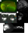
Choroidal metastasis as the first manifestation of renal pelvis carcinoma: A case report
- PMID: 40295273
- PMCID: PMC12040061
- DOI: 10.1097/MD.0000000000042268
Choroidal metastasis as the first manifestation of renal pelvis carcinoma: A case report
Abstract
Rationale: Choroidal metastasis is an uncommon event, especially as the first sign of renal pelvis carcinoma (RPC), a rare subtype of upper tract urothelial carcinoma (UTUC). While the majority of choroidal metastases originate from primary cancers like breast or lung, those arising from urologic cancers are extremely rare. This article describes a case of RPC where the first clinical sign was a choroidal mass.
Patient concerns: A 51-year-old female presented with a 2-week history of decreased vision in her left eye (OS). She reported no prior history of malignancy or significant family history of cancer. Examination revealed retinal detachment and a reddish-white, dome-shaped choroidal lesion. Multimodal imaging, including indocyanine green angiography, Doppler ultrasound, optical coherence tomography confirmed the metastatic nature of the mass.
Diagnoses: Magnetic resonance imaging, computed tomography and whole-body bone scintigraphy with 99mTc-MDP detected multiple metastases to the brain, lungs, and bone, with primary RPC in the left renal pelvis. These findings led to the diagnosis of metastatic RPC.
Interventions: Given the advanced disease stage and poor prognosis, the patient declined invasive treatments such as biopsy or systemic chemotherapy.
Outcomes: Two weeks after diagnosis, the patient succumbed to rapid disease progression.
Lessons: This is a unique case of RPC presenting with tetra-organ metastases involving the lung, bone, brain, and choroid. It underscores the need for a comprehensive systemic evaluation in patients with unexplained choroidal masses, as these may be indicative of an underlying, often asymptomatic, systemic malignancy. Further therapeutic studies are essential to explore effective management strategies and improve outcomes for similar patients with RPC.
Keywords: case report; choroidal metastasis; renal pelvis carcinoma; retinal detachment.
Copyright © 2025 the Author(s). Published by Wolters Kluwer Health, Inc.
Conflict of interest statement
The authors have no conflicts of interest to disclose.
Figures


Similar articles
-
Prescription of Controlled Substances: Benefits and Risks.2025 Jul 6. In: StatPearls [Internet]. Treasure Island (FL): StatPearls Publishing; 2025 Jan–. 2025 Jul 6. In: StatPearls [Internet]. Treasure Island (FL): StatPearls Publishing; 2025 Jan–. PMID: 30726003 Free Books & Documents.
-
Choroidal metastasis as a presenting manifestation of lung cancer: a report of 3 cases and systematic review of the literature.Medicine (Baltimore). 2012 Jul;91(4):179-194. doi: 10.1097/MD.0b013e3182574a0b. Medicine (Baltimore). 2012. PMID: 22732948
-
Contrast-enhanced ultrasound using SonoVue® (sulphur hexafluoride microbubbles) compared with contrast-enhanced computed tomography and contrast-enhanced magnetic resonance imaging for the characterisation of focal liver lesions and detection of liver metastases: a systematic review and cost-effectiveness analysis.Health Technol Assess. 2013 Apr;17(16):1-243. doi: 10.3310/hta17160. Health Technol Assess. 2013. PMID: 23611316 Free PMC article.
-
Are Current Survival Prediction Tools Useful When Treating Subsequent Skeletal-related Events From Bone Metastases?Clin Orthop Relat Res. 2024 Sep 1;482(9):1710-1721. doi: 10.1097/CORR.0000000000003030. Epub 2024 Mar 22. Clin Orthop Relat Res. 2024. PMID: 38517402 Free PMC article.
-
The role of multimodal ophthalmic imaging in diagnosing choroidal metastasis.Int Ophthalmol. 2025 Aug 18;45(1):339. doi: 10.1007/s10792-025-03664-6. Int Ophthalmol. 2025. PMID: 40824326
References
-
- Gontero P, Birtle A, Capoun O, et al. . European association of Urology guidelines on non-muscle-invasive bladder cancer (TaT1 and Carcinoma In Situ)-a summary of the 2024 guidelines update. Eur Urol. 2024;86:531–49. - PubMed
Publication types
MeSH terms
Grants and funding
LinkOut - more resources
Full Text Sources
Medical

