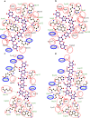
Characterization of two GH10 enzymes with ability to hydrolyze pretreated Sorghum bicolor bagasse
- PMID: 40295346
- PMCID: PMC12037437
- DOI: 10.1007/s00253-025-13484-4
Characterization of two GH10 enzymes with ability to hydrolyze pretreated Sorghum bicolor bagasse
Abstract
In this study, we characterized two novel enzymes of the glycoside hydrolase family 10 (GH10), Xyl10 C and Xyl10E, identified in the termite gut microbiome. The activities of both enzymes were assayed using beechwood xylan, barley β-glucan, and pretreated Sorghum bicolor bagasse (SBB) as substrates. Both enzymes, assessed individually and in combination, showed activity on beechwood xylan and pretreated SBB, whereas Xyl10E also showed activity on barley β-glucan. The composition of pretreated SBB mainly consisted of xylose and arabinose content. Purified Xyl10 C showed optimum xylanase activity in the pH range 7.0-8.0 and at a temperature of 50-60 °C, while Xyl10E was active at a wider pH range (5.0-10.0) and at 50 °C. The residual activities of Xyl10 C and Xyl10E after 8 h of incubation at 40 °C were 85% and 70%, respectively. The enzymatic activity of Xyl10 C increased to 115% in the presence of 5 M NaCl, was only inhibited in the presence of 0.5% sodium dodecyl sulfate (SDS), and decreased with β-mercaptoethanol. The xylanase and glucanase activities of Xyl10E were inhibited only in the presence of MnSO4, NaCl, and SDS. The main hydrolysis enzymatic product of Xyl10 C and Xyl10E on pretreated SBB was xylobiose. In addition, the xylo-oligosaccharides produced by xylanase Xyl10E on pretreated SBB demonstrated promising antioxidant activity. Thus, the hydrolysis products using Xyl10E on pretreated SBB indicate potential for antioxidant activity and other valuable industrial applications. KEY POINTS: • Two novel GH10 xylanases from the termite gut microbiome were characterized. • Xylo-oligosaccharides obtained from sorghum bagasse exhibited antioxidant potential. • Both enzymes and their hydrolysis product have potential to add value to agro-waste.
Keywords: Antioxidant activity of xylo-oligosaccharides; Bifunctional xylanase/β-glucanase; GH10; Pretreated Sorghum bicolor bagasse; Xylanase.
© 2025. The Author(s).
Conflict of interest statement
Declarations. Competing interests: The authors declare no competing interests. Ethical approval: This article does not contain any studies with human participants or animals performed by any of the authors.
Figures


Similar articles
-
Biochemical characterization of the xylan hydrolysis profile of the extracellular endo-xylanase from Geobacillus thermodenitrificans T12.BMC Biotechnol. 2017 May 18;17(1):44. doi: 10.1186/s12896-017-0357-2. BMC Biotechnol. 2017. PMID: 28521816 Free PMC article.
-
Purification and characterization of an endo-xylanase from Trichoderma sp., with xylobiose as the main product from xylan hydrolysis.World J Microbiol Biotechnol. 2019 Oct 31;35(11):171. doi: 10.1007/s11274-019-2747-1. World J Microbiol Biotechnol. 2019. PMID: 31673786
-
Expression and characterization of a novel halophilic GH10 β-1,4-xylanase from Trichoderma asperellum ND-1 and its synergism with a commercial α-L-arabinofuranosidase on arabinoxylan degradation.Int J Biol Macromol. 2024 Dec;282(Pt 2):136885. doi: 10.1016/j.ijbiomac.2024.136885. Epub 2024 Oct 23. Int J Biol Macromol. 2024. PMID: 39454924
-
Identification and Characterization of a Novel, Cold-Adapted d-Xylobiose- and d-Xylose-Releasing Endo-β-1,4-xylanase from an Antarctic Soil Bacterium, Duganella sp. PAMC 27433.Biomolecules. 2021 Apr 30;11(5):680. doi: 10.3390/biom11050680. Biomolecules. 2021. PMID: 33946575 Free PMC article.
-
Identification of a novel glycoside hydrolase family 8 xylanase from Deinococcus geothermalis and its application at low temperatures.Arch Microbiol. 2024 Jun 17;206(7):307. doi: 10.1007/s00203-024-04055-8. Arch Microbiol. 2024. PMID: 38884653
References
-
- Åman P, McNeil M, Franzén L-E, Darvill AG, Albersheim P (1981) Structural elucidation, using H.P.L.C.-M.S. and G.L.C.-M.S., of the acidic polysaccharide secreted by Rhizobium meliloti strain 1021. Carbohydr Res 95(2):263–282 10.1016/S0008-6215(00)85582-2
MeSH terms
Substances
Grants and funding
- MISSIONE 4 COMPONENTE 2, INVESTIMENTO 1.3-D.D. 1551.11-10-2022, PE00000004/European Union Next-Generation EU (PIANO NAZIONALE DI RIPRESA E RESILIENZA (PNRR)
- MISSIONE 4 COMPONENTE 2, INVESTIMENTO 1.3-D.D. 1551.11-10-2022, PE00000004/European Union Next-Generation EU (PIANO NAZIONALE DI RIPRESA E RESILIENZA (PNRR)
- MISSIONE 4 COMPONENTE 2, INVESTIMENTO 1.3-D.D. 1551.11-10-2022, PE00000004/European Union Next-Generation EU (PIANO NAZIONALE DI RIPRESA E RESILIENZA (PNRR)
- (PI 085, 089, 122 and 159)/Instituto Nacional de Tecnología Agropecuaria (INTA)
- (2018-#4149, 2019-#3156, 2020-#3570)/Agencia Nacional de Promoción Científica y Tecnológica (ANPCyT) Proyectos de Investigación Científica y Tecnológica (PICT)
LinkOut - more resources
Full Text Sources

