. 2024 Jul 23;2(1):30.
doi: 10.1038/s44298-024-00041-5.

Identification of amino acids restricting HBV receptor function in porcine NTCP

Affiliations

Identification of amino acids restricting HBV receptor function in porcine NTCP

Samuel D Jeske et al. Npj Viruses. .

Erratum in

Abstract

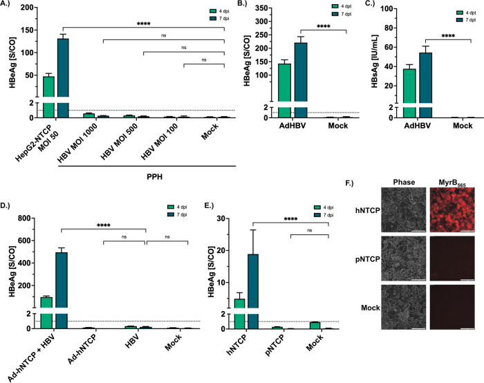
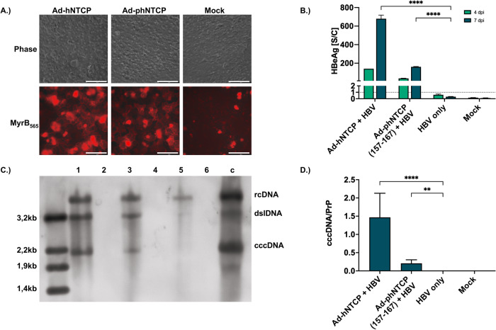
With 254 million chronically infected patients, hepatitis B virus (HBV) continues to be a severe health threat. While animal models play a crucial role in developing new therapies, the availability of preclinical HBV models is very limited. Therefore, novel in vivo infection models are urgently needed. The bona fide HBV receptor, sodium-taurocholate cotransporting polypeptide (NTCP), determines HBV's species and cell-type specificity. Recent studies have indicated that the expression of human NTCP is the only limiting factor for HBV infection in selected species, such as macaques or pigs. Here, we confirm HBV infection of pig hepatocytes expressing human NTCP and show that porcine NTCP does not support HBV binding. By gradually humanizing porcine NTCP and site-directed mutagenesis, we identified amino acids 158 and 167 in porcine NTCP, limiting HBV interaction. In a proof-of-concept experiment, we showed that the expression of porcine NTCP with humanized amino acids 157-167 renders primary porcine hepatocytes fully susceptible to HBV. These results pave the way for generating transgenic pigs with humanized porcine chimeric NTCP as a novel, fully immunocompetent infection model for developing and validating new curative HBV therapies.


Conflict of interest statement

Competing interests: S.D.J., J.M.W., K.F., A.S., and U.P. filed the following patent based on the described findings: Hepatitis B and/or Hepatitis D-permissive cells and animals, International Patent (PCT) application (2019) WO 2019/243 564. All other authors declare no financial or non-financial competing interests.

Figures

Fig. 1
Fig. 1. Expression of hNTCP is the limiting factor for HBV infection in PPH.
AD PPH were isolated from fresh pig liver using the two-step collagenase perfusion method, seeded into 12-well plates, and differentiated with 2% DMSO for 48 h. (A) Differentiated PPH were inoculated with HBV at indicated MOI. PPH not inoculated with HBV served as negative (Mock) and HepG2-NTCP (K7) cells inoculated with HBV (MOI 50 vp/cell) as positive controls. Cell culture medium was collected at days 4 and 7 post-inoculation (dpi) and analyzed for HBeAg. B, C Differentiated PPH were transduced via an adenoviral vector carrying a 1.3-fold HBV genome. Cell culture medium was collected at days 4 and 7 post-transduction (dpt) and analyzed for HBeAg (B) and HBsAg (C). D PPH were transduced with an adenoviral vector expressing hNTCP and subsequently inoculated with HBV at MOI 1000 vp/ml. Cell culture medium was collected at days 4 and 7 post-infection and analyzed for HBeAg. E, F HepG2 cells were transfected with mRNA encoding hNTCP or pNTCP and differentiated with 2% DMSO for 48 h. E Cells were inoculated with HBV (MOI 300 vp/cell), and cell culture medium was collected and analyzed for HBeAg at days 4 and 7 post-infection. F Cells were treated with fluorescently labeled Myrcludex B (MyrB565) and analyzed via bright field (phase contrast) and fluorescence microscopy (scale bar: 100 µm). Representative images are shown. A–E Dotted lines indicate the cut-off between non-reactive and reactive. Experiments were performed in biological triplicates; mean values +/− standard deviation are given. Statistical analysis was performed by one-way ANOVA with Dunnett’s multiple comparison correction (A, D, E) or t-test (B, C). ****p < 0,0001, ns = not significant.
Fig. 2
Fig. 2. Identification of the amino acids to be exchanged in pNTCP for HBV infection.
A Overview of the NTCP aa sequences encoded by the generated mRNA (A1–A6) with a gradual exchange of larger regions of the pNTCP sequence with their human counterparts. B HepG2 cells were transfected with the generated mRNAs (A1–A6), differentiated with 2% DMSO for 48 h, and subsequently inoculated with HBV (MOI 500 vp/cell). Cell culture supernatants were collected at days 4 and 7 post-infection and analyzed for HBeAg. C Overview of the NTCP aa sequences encoded by the generated mRNA (B1-B5) based on pNTCP with aa exchanges in the aa region 155–170. D HepG2 cells were transfected with the generated mRNAs (B1-B5), differentiated with 2% DMSO for 48 h, and subsequently inoculated with HBV (MOI 500 vp/cell). Cell culture supernatants were collected at days 4 and 7 post-infection and analyzed for HBeAg. B, D Experiments were performed in biological triplicates; mean values +/− standard deviation are given. Data were analyzed by one-way ANOVA with Dunnett’s multiple comparison correction. ****p < 0,0001, ns = not significant. B1 = phNTCP (G157K; S158G; I164V; I166V; P167L); B2 = phNTCP (G157K; S158G; I164V; I166V); B3 = phNTCP (S158G; I164V; I166V; P167L); B4 = phNTCP (I164V; I166V; P167L); B5 = phNTCP (S158G; P167L).
Fig. 3
Fig. 3. Generation of stable cell lines expressing hNTCP, pNTCP, and phNTCP variants (157-167) and (S158G; P167L).
HepG2 cell lines stably expressing hNTCP, pNTCP, phNTCP B1 (157-167), and phNTCP B5 (S158G; P167L) were generated. A Cells were treated with fluorescently labeled MyrB565 and analyzed via fluorescence microscopy to investigate HBV binding (scale bar: 100 µm). B Cells were seeded with low density and treated with Atto488-labeled MyrB (MyrB488). Membrane structure was visualized with Alexa Fluor647 labeled wheat germ agglutinin (WGA647), and nuclei were stained with Hoechst33342. High-resolution confocal microscopy was performed to investigate the correct location of the NTCP variants expressed (scale bar: 10 µm). In order to visualize the colocalization of WGA647 and MyrB488, fluorescence intensities along white indicators were compared. C Cells were seeded, differentiated, and inoculated with HBV (MOI 500 vp/cell). Supernatants were collected and analyzed for HBeAg at days 4 and 7 post-infection. The dotted line indicates the cut-off between non-reactive and reactive. Experiments were performed in biological triplicates; mean values +/− standard deviation are given. Data were analyzed by one-way ANOVA with Dunnett’s multiple comparison correction. *p < 0.05, **p < 0.01, ****p < 0.0001.
Fig. 4
Fig. 4. HBV infection of primary porcine hepatocytes with partially humanized porcine NTCP.
PPH were isolated using a 2-step collagenase method and seeded on a 24-well plate. Cells were allowed to attach for 24 h, transduced with adenoviral vectors expressing hNTCP or phNTCP (157-167), and subsequently differentiated in DMSO 2% for 48 h. A Cells were stained with fluorescently labeled Myrcludex B (Myr565) and analyzed via fluorescence microscopy, showing binding to the NTCP variants expressed on the cell surface (scale bars: 100 µm). BD In parallel, cells were inoculated with HBV (MOI 1000 vp/cell). B Cell culture supernatants were collected at days 4 and 7 post-infection and analyzed for HBeAg. C 7 days post-infection, southern blot analysis was performed utilizing a modified Hirt extraction procedure. Analyzed samples include (1) PPH transduced with Ad-hNTCP and inoculated with HBV, (2) PPH transduced with Ad-hNTCP only, (3) PPH transduced with Ad-phNTCP (157-167) and inoculated with HBV, (4) PPH transduced with Ad-phNTCP (157-167) only, (5) naive PPH inoculated with HBV, (6) naive PPH without HBV, (c) HBV infected HepG2-NTCP as a positive control. D 7 days post-infection, cells were lysed, total DNA was isolated from the lysate and analyzed for cccDNA via PCR. Values are displayed in relation to prion protein gene. B, D Mean values +/− standard deviation of at least three experiments are given. Statistical analysis was performed by (B) one-way ANOVA with Dunnett’s multiple comparison correction or (D) Kruskal–Wallis test with Dunn’s multiple comparison correction. **p < 0,01, ****p < 0,0001. For (B), the dotted line indicates the cut-off between non-reactive and reactive.

References

    1. World Health Organization. Global hepatitis report 2024: action for access in low- and middle-income countries. (World Health Organization, 2024). https://iris.who.int/handle/10665/376461.
    1. Lucifora, J. & Protzer, U. Attacking hepatitis B virus cccDNA–The holy grail to hepatitis B cure. J. Hepatol.64, S41–S48 (2016). - PubMed
    1. Gehring, A. J. & Protzer, U. Targeting innate and adaptive immune responses to cure chronic HBV infection. Gastroenterology156, 325–337 (2019). - PubMed
    1. Wettengel, J. M. & Burwitz, B. J. Innovative HBV animal models based on the entry receptor NTCP. Viruses12, 828 (2020). - PMC - PubMed
    1. Institute of Medicine and National Research Council. Chimpanzees in Biomedical and Behavioral Research: Assessing the Necessity. (The National Academies Press, Washington, DC, 2011). 10.17226/13257. - PubMed

LinkOut - more resources