. 2024 May 2;2(1):19.
doi: 10.1038/s44298-024-00027-3.

Human macrophages infected with Egyptian Rousette bat-isolated Marburg virus display inter-individual susceptibility and antiviral responsiveness

Affiliations

Human macrophages infected with Egyptian Rousette bat-isolated Marburg virus display inter-individual susceptibility and antiviral responsiveness

Ivet A Yordanova et al. Npj Viruses. .

Abstract

Marburg virus (MARV) is a highly pathogenic filovirus and a causative agent of sporadic zoonotic viral hemorrhagic fever outbreaks with high case fatality rates. In humans, filoviruses like MARV and Zaire Ebola virus (EBOV) target, among others, innate immune cells like dendritic cells and macrophages (MΦs). Filovirus-infected dendritic cells display impaired maturation and antigen presentation, while MΦs become hyper-activated and secrete proinflammatory cytokines and chemokines. Our current understanding of human macrophage responses to MARV remains limited. Here, we used human monocyte-derived macrophages (moMΦs) to address how their phenotype, transcriptional profile, and protein expression change upon an in vitro infection with a bat isolate of MARV. Confirming its tropism for macrophages, we show that MARV induces notable shifts in their transcription distinct from responses induced by lipopolysaccharide (LPS), marked by upregulated gene expression of several chemokines, type I interferons, and IFN-stimulated genes. MARV infection also elicited pronounced inter-individually different transcriptional programs in moMΦs, the induction of Wnt signaling-associated genes, and the downregulation of multiple biological processes and molecular pathways.


Conflict of interest statement

Competing interests: The authors declare no competing interests.

Figures

Fig. 1
Fig. 1. MARV replication and progeny production in human moMΦs 1 day following MARV-ZsG infection.
a ZsG expression in MARV-infected versus mock-infected human moMΦs, as observed under a fluorescent microscope. b Example FACS plots and c percentages of ZsG+ cells within live CD11b+CD14+ moMΦs in individual donors, quantified using flow cytometry. d Virus production in cell culture supernatants of MARV-infected moMΦs, quantified using a focus assay. The data are pooled from two independent experiments with n = 3–5 donors per experiment.
Fig. 2
Fig. 2. Cell surface marker expression in moMΦs 1-day post-infection with MARV-ZsG.
a Dimensionality reduction analysis (tSNE) of the cell populations found in in vitro-differentiated human moMΦ cultures. b Overlay tSNE plots illustrating the expression patterns of macrophage surface markers CD163, CD206, HLA-DR, and CD40, as well as of ZsG, within MARV-infected moMΦ cultures. c Example histogram plot and median fluorescence intensity (MFI) of the surface expression levels of CD163 in ZsG+ and ZsG- moMΦs. The results in c are pooled from two independent experiments with n = 3–5 donors per experiment. Statistical analysis was done using a Wilcoxon Signed Rank test. **p < 0.01.
Fig. 3
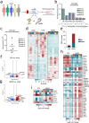
Fig. 3. Differential gene expression in LPS-treated and MARV-infected human moMΦs.
a moMΦs were differentiated from five individual healthy blood donors. Cells from each donor were either mock-infected, LPS-treated, or MARV-infected and were harvested after 1 day of treatment or infection for bulk RNAseq analysis. b Normalized counts of all 7 MARV genes in individual donors, measured using RNA sequencing. c Viral production in cell culture supernatants, quantified using qRT-PCR detection of MARV NP and shown as gene copies per µL cell culture supernatant. d Global gene expression profile of mock-infected, LPS-treated, and MARV-infected moMΦs from individual donors. e Total numbers of significantly upregulated and downregulated DEGs in moMΦs following in vitro LPS or MARV challenge (filtered for treatment group-level p-adjusted values < 0.05). Volcano plots illustrating significant DEGs across all five donors in (f) LPS-treated and (g) MARV-infected cells, compared with mock-infected controls. Genes upregulated twofold or higher are highlighted in red, while genes downregulated twofold or lower are highlighted in blue. h Heatmap illustrating immune-related DEGs in LPS-treated and MARV-infected moMΦs. i Heatmap illustrating Wnt signaling-related DEGs in LPS-treated and MARV-infected moMΦs. The heatmaps in h and i are shown as log2 fold change against mock-infected controls.
Fig. 4
Fig. 4. Baseline global gene expression and cell surface receptor expression in virus non-responders versus responders.
a RNAseq analysis set-up, grouping and comparing MARV non-responder donors 1 and 3 versus responder donors 2, 4, and 5 at baseline (mock-infected control samples). b Global gene expression in individual non-responder versus responder donors. cj Normalized gene counts of genes encoding cell surface receptors associated with filovirus attachment and entry.
Fig. 5
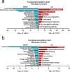
Fig. 5. Functional annotation analysis of DEGs in MARV-infected moMΦs (differentially expressed against mock-infected controls).
a Biological process and b Molecular function gene ontology (GO) terms, enriched among DEGs in MARV-infected moMΦs. GO terms enriched within genes upregulated twofold or higher are shown in red, while genes downregulated twofold or lower are highlighted in blue. Functional annotation analysis was performed using DAVID.
Fig. 6
Fig. 6. Cytokine and chemokine expression in human moMΦs.
Protein levels of a proinflammatory cytokines IFNα, IL-6, and TNF and b chemokines CCL4, CCL5, and CXCL10 in cell culture supernatants of mock-infected, LPS-treated and MARV-infected moMΦs 1 day post-treatment or infection, measured using a 34-plex human Luminex assay. Donors defined as MARV non-responders are marked as triangles, while responder donors are marked in squares. Supernatants from three additional donors (white circles) were included in the Luminex assay. The data are pooled from 2 independent experiments with =3–5 donors per experiment. Statistical analysis was performed using a Kruskall–Wallis multiple comparisons test. ***p < 0.001.

Similar articles

References

    1. Feldmann, H. Marburg hemorrhagic fever—the forgotten cousin strikes. N. Engl. J. Med.355, 866–869 (2006). - PubMed
    1. Brauburger, K., Hume, A. J., Mühlberger, E. & Olejnik, J. Forty-five years of marburg virus research. Viruses4, 1878–1927 (2012). - PMC - PubMed
    1. Srivastava, S. et al. Emergence of Marburg virus: a global perspective on fatal outbreaks and clinical challenges. Front. Microbiol.10.1056/NEJMp068160 (2023). - PMC - PubMed
    1. Gupta, M., Spiropoulou, C. & Rollin, P. E. Ebola virus infection of human PBMCs causes massive death of macrophages, CD4 and CD8 T cell sub-populations in vitro. Virology364, 45–54 (2007). - PubMed
    1. Wanninger, T. G. et al. Macrophage infection, activation, and histopathological findings in ebolavirus infection. Front. Cell Infect. Microbiol.12, 1–17 (2022). - PMC - PubMed

LinkOut - more resources