. 2025 Apr 28;26(1):30.
doi: 10.1186/s12868-025-00947-7.

Body weight-supported treadmill training reduces glial scar overgrowth in SCI rats by decreasing the reactivity of astrocytes during the subacute phase

Affiliations

Body weight-supported treadmill training reduces glial scar overgrowth in SCI rats by decreasing the reactivity of astrocytes during the subacute phase

Jili Cai et al. BMC Neurosci. .

Abstract

Background: Spinal cord injury is followed by glial scar formation, which was long seen mainly as a physical barrier preventing axonal regeneration. Glial scar astrocytes lead to glial scar formation and produce inhibitory factors to prevent axons from growing through the scar, while inhibiting the conversion of reactive astrocytes into glial scar-forming astrocytes may represent an ideal treatment for CNS injury. Exercise is a non-invasive and effective therapeutic intervention for clinical rehabilitation of spinal cord injury. However, its precise therapeutic mechanisms still need to be continuously explored.

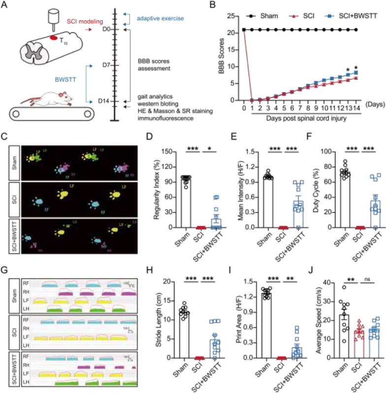
Methods: 30 rats were randomly assigned to three groups (Sham, SCI, SCI + BWSTT; n = 10 rats per group). In this study, we employed the BBB scales and gait analysis system to examine the behavioral functions of the rats in each group. Furthermore, we utilized immunoblotting of spinal cord tissue at the injury site, in addition to histological staining and immunofluorescence staining, to explore glial scar aggregation and axonal regeneration in each group of rats.

Results: Our results revealed that hindlimb motor function was significantly improved in SCI rats after a sustained subacute period of BWSTT, accompanied by the promotion of histological repair and nerve regeneration. Subsequent immunofluorescence staining and immunoblotting showed diminished astrocyte reactivity in the region surrounding the spinal cord injury as well as reduced expression and distribution of collagen fibers near the lesion after BWSTT. Additionally, a significant decrease in the expression of MMP-2/9, which is closely related to astrocyte migration, was observed in the vicinity of spinal cord tissue lesions.

Conclusion: Our study demonstrates that a sustained BWSTT intervention during the subacute phase of spinal cord injury can effectively reduce astrocyte reactivity and glial scarring overgrowth, thereby facilitating functional recovery after SCI.

Keywords: Astrocyte reactivity; Body weight-supported treadmill training; Glial scar; Matrix metalloproteinase-2/9; Spinal cord injury.


Conflict of interest statement

Declarations. Ethics approval and consent to participate: All experiments involving animals were approved by the Ethics Committee for Animal Experiments of Nanjing Medical University (license No. IACUC-2206045 and No. IACUC-2403019), Jiangsu Province, China. Consent for publication: Not applicable. Competing interests: The authors declare no competing interests.

Figures

Fig.1
Fig.1
BWSTT improved functional recovery after SCI. A Time-line diagram of spinal cord injury, BWSTT treatment, and experimental analysis in rats. B The BBB scores in each group (n = 10). CJ The CatWalk XT® automated gait analysis results in each group (n = 10). The representative images of footprint intensity during a pedestrian cycle C and footprint pressure–time chart G in each group. The quantification analysis of the CatWalk of gait analysis parameters in each group, D regularity index, E mean intensity (H/F), F duty cycle, H stride length, I print area, J average speed. Values are expressed as the mean ± s.e.m, ⋆p < 0.05, ⋆⋆p < 0.01, and ⋆⋆⋆p < 0.001, as determined by one-way ANOVA. RF-right forelimb, RH-right hindlimb, LF-left forelimb, LH-left hindlimb
Fig. 2
Fig. 2
BWSTT promotes histological repair of the damaged spinal cord. A Representative H&E staining and Masson staining images in longitudinal sections of the spinal cord from each group (n = 3). The line labels the injured area and * Indicates the lesion center. Scale bar = 2000 μm, 1000 μm. B The schematic representation of the injury site is utilized for pathological staining. C, D Quantification analysis of the injured area percentage of the H&E staining (C) and collagen volume fraction of the Masson staining (D). Values are expressed as the mean ± s.e.m, * p < 0.05 as determined by one-way ANOVA
Fig. 3
Fig. 3
BWSTT inhibits neuronal cell death and promotes axonal regeneration after SCI. A Representative immunofluorescence images of NeuN (a neuronal marker) (green) from the anterior horn area of the spinal cord (transverse sections of the caudal region of the injured spinal cord) in each group following SCI (n = 3). Nuclei were counterstained with DAPI (blue). Scale bar = 100 μm, 50 μm. B, C Quantification of the number of NeuN+ cells/mm2 in (A) and NF200+ fluorescence intensity area in (D). D Representative immunofluorescence images of NF200 (key components of the neuronal cytoskeleton) (green) and DAPI (blue) in longitudinal sections of the spinal cord from each group after SCI (n = 3). Scale bar = 500 μm and * Indicates lesion center. Values are expressed as the mean ± s.e.m, * p < 0.05, ** p < 0.01, and *** p < 0.001, as determined by one-way ANOVA
Fig. 4
Fig. 4
BWSTT reduces the astrocyte reactivity and the expression of collagen I. A Representative immunofluorescence images of GFAP (red) and DAPI (blue) of the injured site in longitudinal sections of the spinal cord form each group (n = 3). Scale bar = 100 μm, and * Indicates the lesion center. B, C Quantification analysis of the GFAP fluorescence intensity B and the GFAP+ fluorescence area in the cross sections in each group. DF Representative images of Western blotting and quantification analysis of collagen I, vimentin, and β-actin in each group (n = 4). G Representative images of Picrosirius Red staining in longitudinal sections of the spinal cord form each group (n = 3). Scale bar = 1000 μm, and * Indicates the lesion center. H Quantification analysis of the collagen area of the injured site in (G). Values are expressed as the mean ± s.e.m, *p < 0.05, **p < 0.01, and ***p < 0.001, as determined by one-way ANOVA
Fig. 5
Fig. 5
BWSTT downregulates the Matrix Metalloproteinase-2/9(MMP-2/9) after SCI. A Representative images of Western blotting of MMP-9, MMP-2, and β-actin in each group after SCI (n = 4). B, C Quantification analysis of MMP-9 (B) and MMP-2 (C) relative protein expression in the epicenter. Values are expressed as the mean ± s.e.m, *p < 0.05, **p < 0.01, and ***p < 0.001, as determined by one-way ANOVA

Similar articles

References

    1. Crispo JAG, Kuramoto LK, Cragg JJ. Global burden of spinal cord injury: future directions. Lancet Neurol. 2023;22:976–8. - PubMed
    1. Zheng B, Tuszynski MH. Regulation of axonal regeneration after mammalian spinal cord injury. Nat Rev Mol Cell Biol. 2023;24:396–413. - PubMed
    1. Gong L, Gu Y, Han X, Luan C, Liu C, Wang X, et al. Spatiotemporal dynamics of the molecular expression pattern and intercellular interactions in the glial scar response to spinal cord injury. Neurosci Bull. 2023;39:213–44. - PMC - PubMed
    1. Lima R, Monteiro A, Salgado AJ, Monteiro S, Silva NA. Pathophysiology and therapeutic approaches for spinal cord injury. Int J Mol Sci. 2022;23:13833. - PMC - PubMed
    1. Clifford T, Finkel Z, Rodriguez B, Joseph A, Cai L. Current advancements in spinal cord injury research—glial scar formation and neural regeneration. Cells. 2023;12:853. - PMC - PubMed

MeSH terms

LinkOut - more resources