

Neurologic symptoms as a hallmark of glymphatic alteration in recovered patients with COVID-19
- PMID: 40295959
- PMCID: PMC12036239
- DOI: 10.1186/s12883-025-04198-1
Neurologic symptoms as a hallmark of glymphatic alteration in recovered patients with COVID-19
Abstract
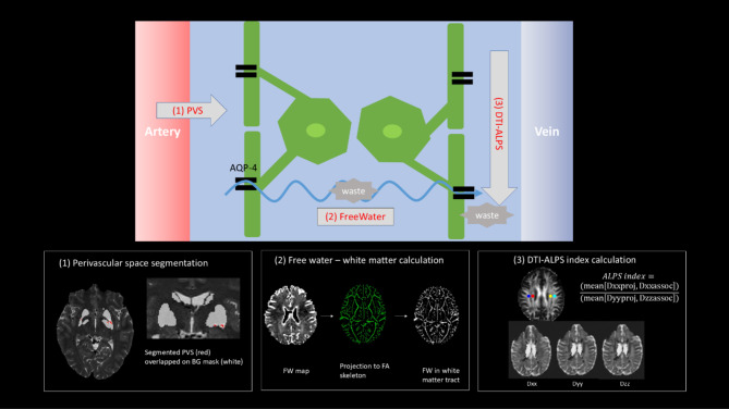
Background: The glymphatic system is a glial-based perivascular network that facilitates the clearance of metabolic waste from the brain. Dysfunction of the glymphatic system, along with neurological symptoms such as cognitive deficits and olfactory dysfunction, has been reported in patients with coronavirus disease (COVID-19). However, the link between these neurological symptoms and alterations in the glymphatic system remains unclear. In this study, we aimed to evaluate magnetic resonance imaging (MRI)-based measures of the glymphatic system in patients recovered from COVID-19 with and without neurological symptoms.
Methods: This study included 89 patients who recovered from respiratory infections, of whom 71 had confirmed COVID-19 (20 experienced anosmia and 41 had cognitive symptoms). Three MRI-based measures were quantified and compared: the dilated perivascular spaces (dPVS), free water (FW) fraction, and diffusion tensor image analysis along the perivascular spaces (DTI-ALPS). A partial correlation network was used to assess the relationships between COVID-19 infection, neurological symptoms, and glymphatic measures.
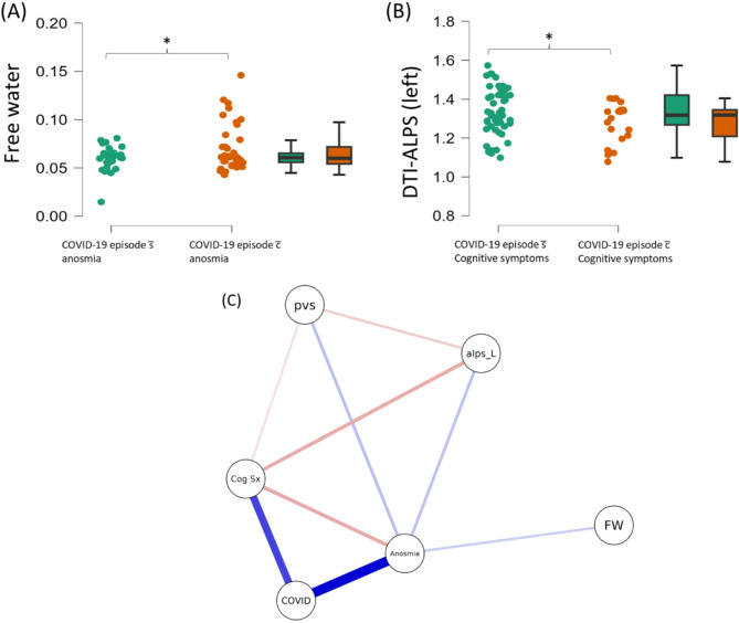
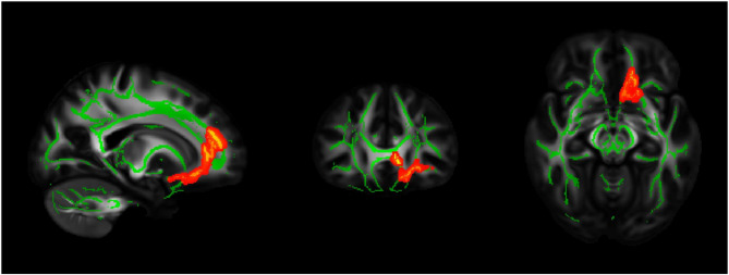
Results: COVID-19 patients with anosmia had increased FW in the left orbitofrontal area compared to those without anosmia (mean difference: 0.01, p = 0.48), while patients with cognitive symptoms showed decreased left-sided DTI-ALPS (mean difference: 0.06, p = 0.40). Neurological symptoms mediate the relationship between COVID-19 and glymphatic system measures.
Conclusions: Our findings imply that neurological symptoms accompanied by COVID-19 are linked to distinct alterations in the glymphatic system, suggesting a potential association between neuroinvasion and neuroinflammatory processes related to COVID-19.
Keywords: Anosmia; COVID-19; DTI-ALPS; Glymphatic alteration.
© 2025. The Author(s).
Conflict of interest statement
Declarations. Ethics approval and consent to participate: All participants gave their informed consent, and the Ethics Committee of Clínica Alemana—Universidad del Desarrollo approved all experimental procedures. The consent process and all experimental methods adhered to Chilean national laws, institutional policies, and the Declaration of Helsinki, and have any necessary ethics permissions to share the data publicly under a Creative Commons CC0 license ( https://docs.openneuro.org/faq.html ). Consent for publication: Not applicable. Competing interests: The authors declare no competing interests.
Figures
Similar articles
-
Asymmetrical glymphatic dysfunction in patients with long Covid associated neurocognitive impairment- correlation with BBB disruption.BMC Neurol. 2025 Mar 19;25(1):112. doi: 10.1186/s12883-025-04133-4. BMC Neurol. 2025. PMID: 40108491 Free PMC article.
-
Neuroimaging findings related to glymphatic system alterations in older adults with metabolic syndrome.Neurobiol Dis. 2023 Feb;177:105990. doi: 10.1016/j.nbd.2023.105990. Epub 2023 Jan 5. Neurobiol Dis. 2023. PMID: 36621631
-
A "glympse" into neurodegeneration: Diffusion MRI and cerebrospinal fluid aquaporin-4 for the assessment of glymphatic system in Alzheimer's disease and other dementias.Hum Brain Mapp. 2024 Aug 15;45(12):e26805. doi: 10.1002/hbm.26805. Hum Brain Mapp. 2024. PMID: 39185685 Free PMC article.
-
Emerging non-invasive MRI techniques for glymphatic system assessment in neurodegenerative disease.J Neuroradiol. 2025 May;52(3):101322. doi: 10.1016/j.neurad.2025.101322. Epub 2025 Jan 31. J Neuroradiol. 2025. PMID: 39894249 Review.
-
Diffusion Tensor Image Analysis ALong the Perivascular Space (DTI-ALPS): Revisiting the Meaning and Significance of the Method.Magn Reson Med Sci. 2024 Jul 1;23(3):268-290. doi: 10.2463/mrms.rev.2023-0175. Epub 2024 Apr 2. Magn Reson Med Sci. 2024. PMID: 38569866 Free PMC article. Review.
References
-
- Organization WH. A clinical case definition of post COVID-19 condition by a Delphi consensus, 6 October 2021. In.: World Health Organization; 2021.
-
- Li H, Jacob MA, Cai M, Kessels RP, Norris DG, Duering M, De Leeuw F-E, Tuladhar AM. Perivascular spaces, diffusivity along perivascular spaces, and free water in cerebral small vessel disease. Neurology. 2024;102(9):e209306. - PubMed
MeSH terms
Grants and funding
LinkOut - more resources
Full Text Sources
Medical
