

Evaluation of accuracy of three-dimensional printing and three-dimensional miniplates in treatment of anterior mandibular fractures: a prospective clinical study
- PMID: 40296061
- PMCID: PMC12038927
- DOI: 10.1186/s12903-025-05935-1
Evaluation of accuracy of three-dimensional printing and three-dimensional miniplates in treatment of anterior mandibular fractures: a prospective clinical study
Abstract
Background: There are strong torsional forces acting on the anterior mandible fractures. Maxillofacial surgery makes extensive use of digital technology, and three-dimensional printing is now an integral element of the workflow in several areas of oral and maxillofacial surgery. Three-dimensional (3D) miniplates had been proposed by several researchers as a good option for mandibular fracture fixation. This clinical trial was conducted to evaluate the accuracy of virtual planning in anterior mandibular fixation by comparing postoperative outcomes with preoperative virtual planning and to evaluate bone healing by measuring bone density after using pre-bent 3D miniplates on a 3D model to complete a 3D workflow and to fix the mandible in 3D.
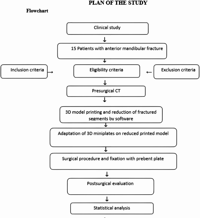
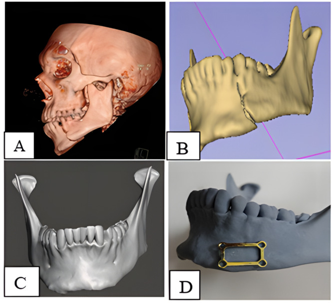
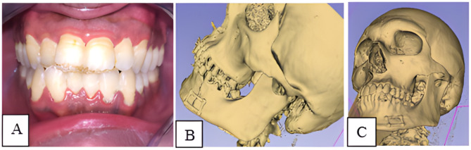
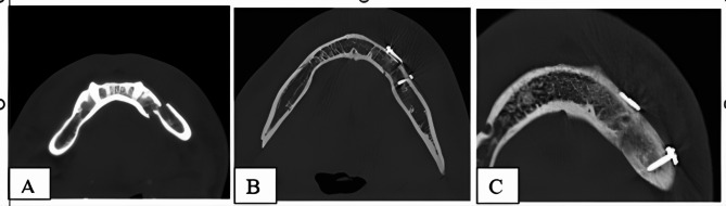
Methods: 15 Patients with anterior mandibular fractures were included in the study. All patients underwent computer tomography (CT) scan, and the data were imported into Mimics software. The unaffected healthy side was mirrored to the fractured side. Bone fixation three-dimensional plates were prebent and adapted on the model printed by the three-dimensional printing machine, submitted to sterilization, and were used for bone reduction and fixation. An immediate postoperative CT scan was taken to evaluate the accuracy of virtual planning and after 3 months for evaluation of bone healing.
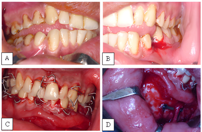
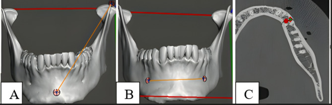
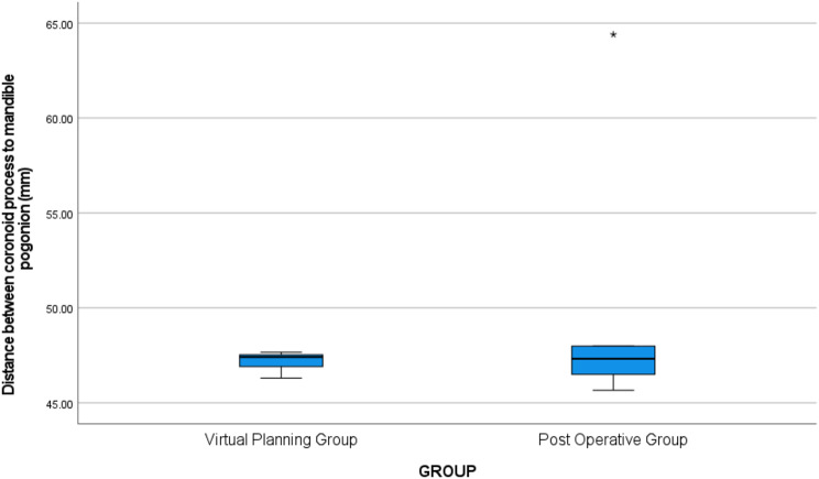
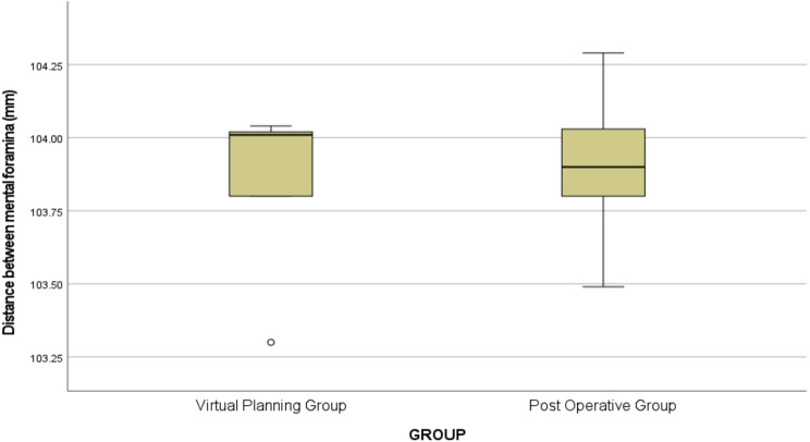
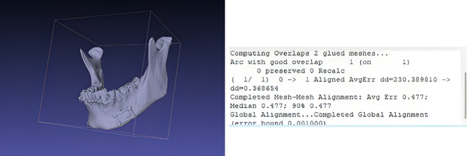
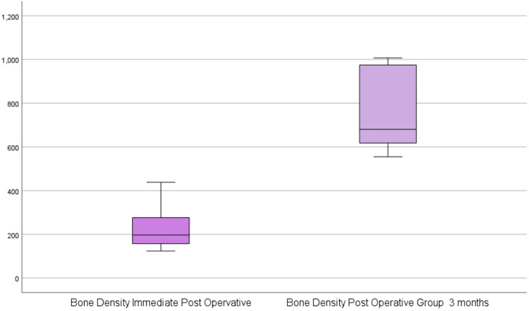
Results: Clinical observation revealed good stable occlusion, and there was no significant difference between the postoperative three-dimensional image of the mandible and the virtually reduced mandible in the preoperative plan, as there was no significant difference between the length and width between anatomical landmarks of the virtually reduced mandible and the postoperative plan (p > 0.05). The bone density measured by Hounsfield units (HU) measured on CT images after 3 months revealed good bone healing as compared to immediate postoperative values (P value < 0.05).
Conclusion: Digital workflow provides an accurate method for the reduction and fixation of anterior mandibular fractures. Also, 3D miniplates provide a good option for symphyseal and parasymphyseal fractures despite their limitations, as in some cases, like comminuted fractures and fractures in and around the mental foramen.
Trial registration: This clinical trial was registered at Alexandria university, on 02/11/2021 under the registration number 0308 - 10/2021. All procedures involving human participants were performed in accordance with Research Ethics Committee, Faculty of Dentistry, Alexandria University under IRB No 00010556 and IORG No 0008839. The current study was retrospectively registered at ClinicalTrials.gov with the identification number NCT06898736 on 27/3/2025. However, all study protocols were predefined, with no deviations from the original methodology.
Keywords: 3D miniplates; Mandibular fracture; Virtual planning. 3D printing.
© 2025. The Author(s).
Conflict of interest statement
Declarations. Ethical approval: This study was performed in accordance with relevant guidelines and regulations. Ethical approval was obtained from Research Ethics Committee, Faculty of Dentistry, Alexandria University with the number 0308 − 10/2021. It was set up and reported according to the CONSORT guidelines. Before starting the study, objectives, risks, and benefits were explained to the patients, and they received both oral and written information about the benefits and risks of the interventions. Signed, informed consent was obtained from all participants before their inclusion in the study. Consent for publication: Not applicable. Competing interests: The authors declare no competing interests.
Figures
References
Publication types
MeSH terms
Associated data
LinkOut - more resources
Full Text Sources
Medical
