. 2025 Aug 15;157(4):760-772.
doi: 10.1002/ijc.35443. Epub 2025 Apr 28.

Patient-derived tumor organoids highlight the potential of precision medicine in managing pancreatic ductal adenocarcinoma

Affiliations

Patient-derived tumor organoids highlight the potential of precision medicine in managing pancreatic ductal adenocarcinoma

Christine Nitschke et al. Int J Cancer. .

Abstract

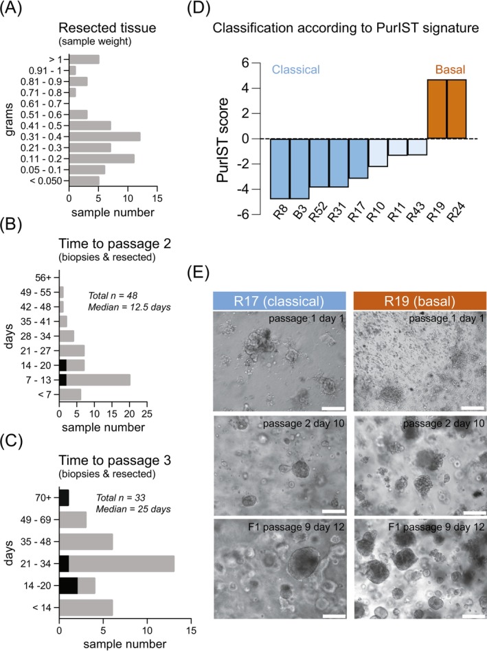
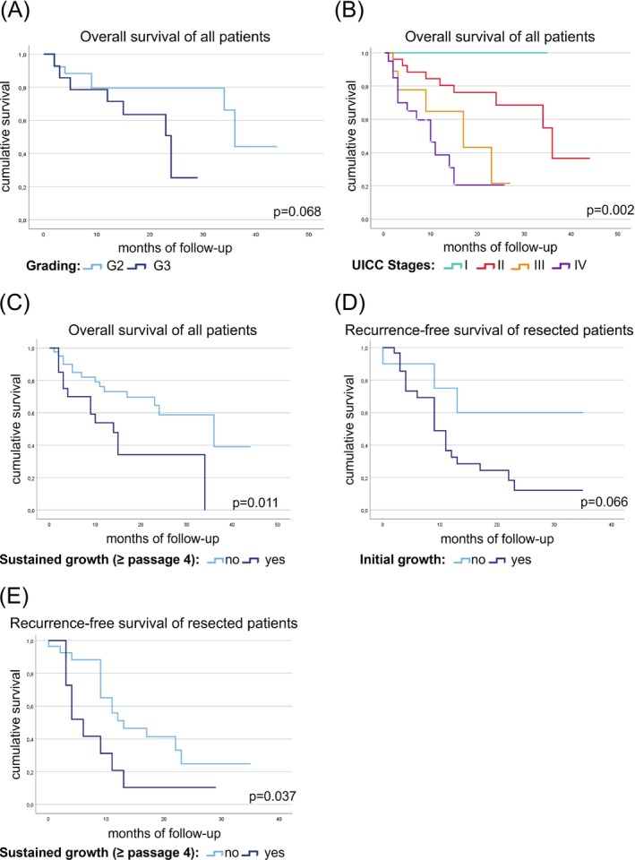
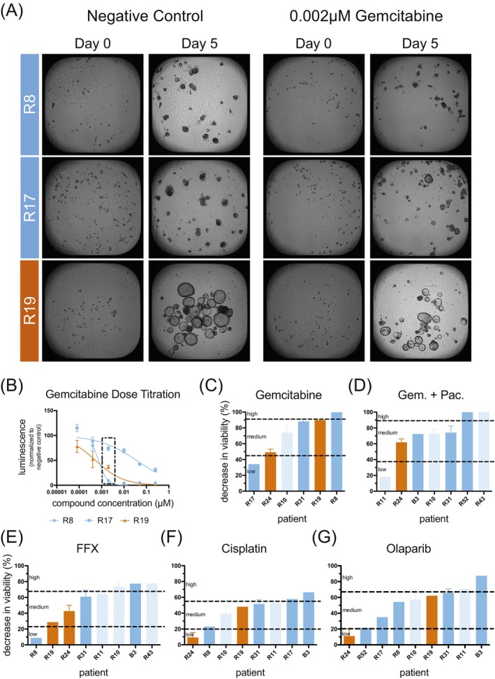
Pancreatic ductal adenocarcinoma (PDAC) ranks among the most lethal cancers, with only 20% of patients qualifying for curative treatment at diagnosis. Three-dimensional tumor organoids capturing patient-specific features of PDAC serve as a valuable disease model. We employed this technology to assess drug sensitivities of patient-derived tumor organoids to clinically relevant drugs and combinations, evaluated culture success rates, and correlated in vitro data with clinicopathological and follow-up information. Tumor organoid cultures were established from PDAC patients undergoing surgical resection (or liver biopsy) and follow-up at a single medical center. Patient-derived cultures displaying sustained growth were analyzed regarding their molecular subtype and utilized for functional drug sensitivity testing (f-DST). Correlative analyses of our PDAC patient cohort (n = 67; n = 42 patients with curative tumor resection and n = 25 palliative patients) revealed a link between tumor organoid growth and reduced patient survival. Furthermore, drug sensitivity profiles (obtained of 10 patient-derived cultures) revealed notable inter-individual differences and mirrored clinical responses to administered drug therapies. f-DST was applicable across tumor organoid cultures of both classical and basal subtype, according to the Purity Independent Subtyping of Tumors (PurIST) classifier. This pilot study confirms the feasibility of deriving and maintaining tumor organoid cultures from heterogeneous samples. Cultures displaying sustained proliferation correlated positively with advanced-stage tumors (Tumour, Node, Metastasis (UICC) stages III and IV). Individual patient case analyses integrating in vitro drug sensitivity profiles with clinical follow-up data suggest that f-DST using tumor organoids could guide future therapeutic strategies. In summary, tumor organoids offer insights into patient-specific responses to treatment, highlighting the potential of precision medicine in managing this challenging cancer.

Keywords: drug testing; pancreatic cancer; precision medicine; tumor organoids.


Conflict of interest statement

Employment or Leadership: Chief Technology Officer of 2cureX (Jacob Thastrup). Stock Ownership: Ownership of 2cureX stocks (Jacob Thastrup, Jürgen Kupper). All other authors declare no conflicts of interest.

Figures

FIGURE 1
FIGURE 1
Characteristics of collected samples and derived tumor organoid cultures. (A) Sample weight of resected tissues varied from below 0.05 g to more than 1 g, with a median sample weight of 0.31 g (distribution shown). (B) Tumor organoid cultures of 48 patients could be established, reaching passage 2 with a median of 12.5 days (distribution is shown, biopsies indicated in red). (C) Tumor organoid cultures of 33 patients could further be expanded, reaching passage 3 with a median of 25 days since preparation (distribution is shown, biopsies indicated in red). (D) Classification according to PurIST signature. (E) Representative bright field images are shown of two different tumor organoid cultures of different subtypes, as determined by PurIST. Light and dark blue = classical subtype, orange = basal subtype. Scale bar = 200 μm. Brightness and contrast of bright field images were enhanced for visualization purposes. Original picture files were submitted to the journal and are available upon reasonable request.
FIGURE 2
FIGURE 2
Kaplan–Meier plots. Data depict overall survival (OS) of pancreatic cancer patients (n = 62) in relation to (A) grading, (B) UICC stages, and (C) sustained growth of patient‐derived tumor organoid cultures. Five patients with a Clavien‐Dindo grade of 5 were excluded from OS analyses. Additional plots depict recurrence‐free survival (RFS) of pancreatic cancer patients operated with curative intent (n = 42) in relation to (D) initial establishment of patient‐derived tumor organoids (passage #2 reached) and (E) sustained growth of tumor organoid cultures.
FIGURE 3
FIGURE 3
Functional drug sensitivity testing of tumor organoids. (A) Representative brightfield images of three different tumor organoid cultures, representing different subtypes, cultured to a size of 40–70 μm, seeded onto a 384‐well microarray plate pre‐loaded with Matrigel® and settled to enable microscopic analysis. Where indicated, cultures were supplemented with Gemcitabine and tumor organoid growth/growth stagnation/disintegration was followed until Day 5. (B) Endpoint analysis using the CellTiter‐Glo® 3D Cell Viability Assay was performed to record dose–response curves for Gemcitabine (n = 5 technical replicates/data point) of three tumor organoid cultures. (C) Using IndiTreat® Single Concentration assay plates, Gemcitabine sensitivity data were acquired for additional cultures via CellTiter‐Glo® endpoint analysis. The decrease in viability relative to the control was extrapolated from dose‐titration curves (no standard error plotted) or IndiTreat® Single Concentration screens (n = 6 technical replicates). Sensitivity categories were assigned by calculating the mean of the data set and placing the dividers at mean ± standard deviation to structure the cohort. Similarly, compiled functional drug sensitivity testing data for (D) Gemcitabine + Paclitaxel (Gem. + Pac.), (E) FOLFIRINOX (FFX), (F) Cisplatin and (G) Olaparib are shown. Light and dark blue = classical subtype, orange = basal subtype.

Similar articles

References

    1. Huang L, Holtzinger A, Jagan I, et al. Ductal pancreatic cancer modeling and drug screening using human pluripotent stem cell‐ and patient‐derived tumor organoids. Nat Med. 2015;21(11):1364‐1371. doi: 10.1038/nm.3973 - DOI - PMC - PubMed
    1. Driehuis E, van Hoeck A, Moore K, et al. Pancreatic cancer organoids recapitulate disease and allow personalized drug screening. Proc Natl Acad Sci U S A. 2019;116(52):26580‐26590. doi: 10.1073/pnas.1911273116 - DOI - PMC - PubMed
    1. Mazur R, Trna J. Principles of palliative and supportive Care in Pancreatic Cancer: a review. Biomedicine. 2023;11(10):2690. doi: 10.3390/biomedicines11102690 - DOI - PMC - PubMed
    1. Garcea G, Dennison AR, Pattenden CJ, Neal CP, Sutton CD, Berry DP. Survival following curative resection for pancreatic ductal adenocarcinoma. A systematic review of the literature. JOP. 2008;9(2):99‐132. - PubMed
    1. Riedl JM, Posch F, Horvath L, et al. Gemcitabine/nab‐paclitaxel versus FOLFIRINOX for palliative first‐line treatment of advanced pancreatic cancer: a propensity score analysis. Eur J Cancer. 2021;151:3‐13. doi: 10.1016/j.ejca.2021.03.040 - DOI - PubMed