. 2025 Apr 18;33(5):1173-1187.
doi: 10.32604/or.2025.057842. eCollection 2025.

Mycobacterial antigen Ag85B restrains Hodgkin lymphoma tumor growth by inhibiting autophagy

Affiliations

Mycobacterial antigen Ag85B restrains Hodgkin lymphoma tumor growth by inhibiting autophagy

Yongfeng Cheng et al. Oncol Res. .

Abstract

Background: The growth of the B-cell lymphoma subtype, Hodgkin lymphoma (HL), is associated with increased autophagy. A mycobacterial antigen, Ag85, has been reported to inhibit cell autophagy under a variety of conditions. Whether Ag85 could inhibit autophagy in HL is unknown.

Methods: Lymph node samples from patients with HL and healthy controls were collected to assess proliferation and autophagy. The human HL cell line, L-428, was cultured and subjected to Ag85B treatment. Autophagy in L-428 cells was evaluated through western blotting analysis, immunohistochemistry, and transmission electron microscopy. Apoptosis in these cells was measured using flow cytometry and western blotting. The associated signaling pathways were also analyzed utilizing western blotting. The in vivo impact of Ag85B was studied using BALB/c Nude mice xenografted with L-428 cells.

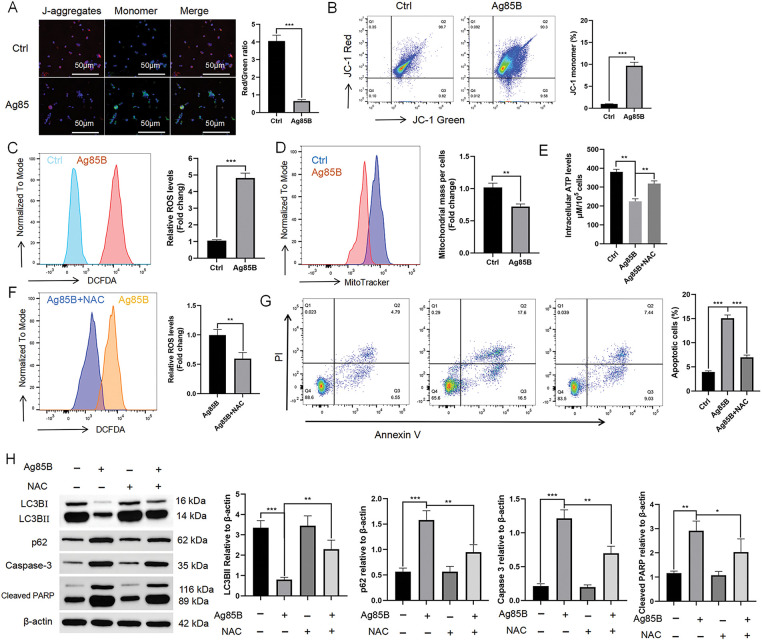
Results: We observed increased proliferation and autophagy in primary lymphoma tissues of patients. Administration of Ag85B inhibited the proliferation and autophagy of HL cell lines. Moreover, Ag85B promoted apoptotic pathway activation in vitro, which might be associated with mitochondrial dysfunction. Mechanistically, Ag85B inhibits autophagy by activating the phosphatidylinositol-4,5-bisphosphate 3-kinase/protein kinase B/mechanistic target of rapamycin kinase (PI3K/AKT/mTOR) and mitogen-activated protein kinase (MAPK) pathways. Ag85B also inhibited lymphoma growth in mice xenografted with HL cell lines, but no potential toxicity was observed.

Conclusion: Altogether, these results suggest that Ag85B inhibits HL growth via autophagy regulation. Current treatments for HL are associated with adverse events; therefore, Ag85B-mediated autophagy inhibition might be a promising strategy in to treat HL.

Keywords: Autophagy; Hodgkin disease; Mitogen-activated protein kinases; Mycobacterial antigen; Protein kinase B.


Conflict of interest statement

The authors declare no conflicts of interest to report regarding the present study.

Figures

Figure 1
Figure 1. Proliferation and autophagy are increased in HL. A. Ki-67 staining of lymphoma primary tissue (patients with HL) compared with normal lymphoid tissue (non-malignant tumor patients). Scale bars = 20 μm. B. The levels of LC3B, Atg14, Beclin-1, and p62 in lymphoma primary tissue compared with those in normal lymphoid tissue were assessed employing western blotting. Right panel: densitometry analysis. C. Immunohistochemical staining of ULK1/2, Park2, PINK-1, Lamp1/2, and Beclin-1 in lymphoma primary tissue and normal lymphoid tissue. Scale bar = 50 μm. Data are expressed as the mean ± SEM from two independent experiments (n = 6). *p < 0.05, **p < 0.01, ***p < 0.001. Ctrl, control; HL, Hodgkin lymphoma; Ki-67, marker of proliferation Ki-67; LC3B, microtubule associated protein 1 light chain 3 beta; Atg14, autophagy related 14; p62, sequestosome 1; lamp, lysosomal associated membrane protein; PINK-1, PTEN induced kinase 1; parkin RBR E3 ubiquitin protein ligase, Park2; ULK, Unc-51 like autophagy activating kinase.
Figure 2
Figure 2. Ag85B inhibits autophagy in L-428 cells. A. Levels of LC3B and p62 proteins measured using western blotting in L-428 cells treated with Ag85B or vehicle. B. Western blotting for the levels of Beclin-1 and Atg14 in L-428 cells administered with Ag85B or vehicle. C. Fluorescence microscopy was utilized to detect the level of mRFP+/GFP+(yellow) and mRFP+/GFP-(red) LC3 puncta in HL cells treated/not treated with Ag85B (4 μg/mL) for 24 h. Typical images and fluorescent LC3 puncta quantitative analysis are shown. Scale bars = 20 μm. D. Immunoblotting assessment of the levels of p62 and LC3 in lysates from L-428 cells treated/not treated withAg85B for 24 h, with or without E64d and PepA (both at 10 μg/mL; sigma). E. TEM observation of autophagosomes in L-428 cells treated/not treated with Ag85B. The autophagosome is shown by an arrow. Scale bars = 5 μm. Data are shown as the mean ± SEM from 2–3 independent tests (n = 6). **p < 0.01, ***p < 0.001, NS, no significance. Ctrl, control; HL, Hodgkin lymphoma; Atg14, autophagy related 14; p62, sequestosome 1; RFP, red fluorescent protein; LC3, microtubule associated protein 1 light chain 3; E64d, cysteine protease inhibitor; PepA, pepstatin A; TEM, transmission electron microscopy.
Figure 3
Figure 3. Ag85B inhibits HL cell proliferation by preventing the cell cycle. A. Viability of L-428 cells treated with different doses of Ag85B for 24 h, and detected via a CCK-8 assay. B. Ki-67 staining in L-428 cells. Scale bars = 20 μm. C. A colony formation assay was employed to assess cell proliferation. Scale bars = 100 μm. D. Flow cytometry detection of the progression of the cell cycle in L-428 cells. E, F. Immunoblotting detection of p21, c-MYC, CDK, and cyclin levels in L-428 cells. Data represent the mean ± SEM from 2–3 independent experiments (n = 6). **p < 0.01, ***p < 0.001. Ctrl, control; HL, Hodgkin lymphoma; CCK-8, cell counting kit 8; Ki-67, marker of proliferation Ki-67; CDK, cyclin dependent kinase; c-MYC, MYC proto-oncogene, BHLH transcription factor; p21, cyclin dependent kinase inhibitor 1A; p53, tumor protein p53.
Figure 4
Figure 4. Ag85B induces cells apoptosis. A. Apoptosis in L-428 cells as assessed using flow cytometry. The percentage of Annexin+/PI- cells represents apoptotic cells; Annexin+/PI+ represents late apoptotic or necrotic cells. B, C. Levels of apoptosis-associated proteins were measured via immunoblotting in L-428 cells. Data represent the mean ± SEM from 2–3 independent experiments (n = 6). **p < 0.01, ***p < 0.001. Ctrl, control; PI, propidium iodide; PARP, poly(ADP-ribose) polymerase; DR5, death receptor 5; Bax, Bcl2 associated X; PUMA, p53 up-regulated modulator of apoptosis; Bcl-xl, Bcl-2-like protein 1.
Figure 5
Figure 5. Ag85B-induced autophagy inhibition and apoptosis are associated with mitochondrial reactive oxygen species (ROS) generation. A. Fluorescent probe JC-1 staining of L-428 cells was followed by detection of the membrane potential (MMP) via fluorescence microscopy. Scale bars = 50 μm. B. Flow cytometry detection of the MMP. The histogram shows the percentage of the JC-1 monomers. C. Ag85B was used to treat L-428 cells for 24 h, followed by incubation with 20 μM dichloro-dihydro-fluorescein diacetate (DCFDA) for 30 min, and then the fluorescent intensity was measured. The histograms show the ROS-related mean fluorescence intensity. D. Mitochondrial mass was detected via MitoTracker™ Green FM. E. ATP levels in L-428 cells. F. Flow cytometry detection of ROS levels in L-428 cells treated/not treated with N-acetylcysteine (NAC) at 10 mM. G. Flow cytometry detection of apoptosis in L-428 cells under the indicated conditions. H. Immunoblotting detection of autophagy- and apoptosis-related proteins in L-428 cells. Data represent the mean ± SEM from 2–3 independent experiments (n = 6). *p < 0.05, **p < 0.01, ***p < 0.001. Ctrl, control; LC3B, microtubule associated protein 1 light chain 3 beta; p62, sequestosome 1; PARP, poly (ADP-ribose) polymerase.
Figure 6
Figure 6. Ag85B activates MAPK and PI3K/AKT pathways. A. Following Ag85B treatment of L-428 for 24 h, immunoblotting was employed to detect PI3K, AKT, mTOR, and their phosphorylated forms (p). B. Immunoblotting detection of the MAPK signaling pathway member protein levels after Ag85B treatment. C, D. L-428 cells were incubated with LY294002 (10 μM) or U0126 (10 μM) for 2 h, and then exposed to Ag85 for 24 h. Western blotting was used to detect the phosphorylation of mTOR and ERK1/2. E. Immunoblotting detection of apoptotic and autophagic marker proteins under the indicated conditions. Data represent the mean ± SEM from two to three independent tests (n = 6). *p < 0.05, **p < 0.01, ***p < 0.001. Ctrl, control; MAPK, mitogen activated protein kinase; PI3K, phosphatidylinositol-4,5-bisphosphate 3-kinase; AKT, protein kinase B; mTOR, mechanistic target of rapamycin kinase; ERK, extracellular regulated kinase; p38, mitogen-activated protein kinase 14; GSK3β, glycogen synthase kinase 3 beta; LC3B, microtubule associated protein 1 light chain 3 beta; p62, sequestosome 1; PARP, poly (ADP-ribose) polymerase.
Figure 7
Figure 7. Ag85B inhibits the tumor growth in BALB/c Nude mice xenografted with L-428 cells. A. The body weights of mice under the indicated conditions. B. The tumor volume was detected to draw tumor growth curves. C. Measurement of tumor weights in the different groups. D. H&E staining for the histological evaluation of vital organs. Scale bars = 50 μm. E. Representative immunofluorescence images of Ki-67 staining of tumor samples. Scale bars = 50 μm. F. Representative immunohistochemical images of LC3B, p62, and cleaved caspase-3 staining of tumor samples. Scale bars = 50 μm. G. Apoptotic tumor cells were detected by TUNEL staining. Scale bars = 20 μm. Apoptotic cell numbers were assessed utilizing ImageJ software. Data are shown as the mean ± SEM from two to three independent tests (n = 6). **p < 0.01, ***p < 0.001. Ctrl, control; Ki-67, marker of proliferation Ki-67; LC3B, microtubule associated protein 1 light chain 3 beta; p62, sequestosome 1.

Similar articles

References

    1. Vardiman JW. The world health organization (WHO) classification of tumors of the hematopoietic and lymphoid tissues: an overview with emphasis on the myeloid neoplasms. Chem Biol Interact. 2010;184(1–2):16–20. doi:10.1016/j.cbi.2009.10.009; - DOI - PubMed
    1. Kahn J, Dabaja B, Wu S, Kelly K, Berkahn L, Pavlovsky A, et al. . Classic Hodgkin lymphoma. Hematol Oncol. 2024;42(6):e3239. doi:10.1002/hon.3239; - DOI - PubMed
    1. Weniger MA, Küppers R. Molecular biology of Hodgkin lymphoma. Leukemia. 2021;35(4):968–81. doi:10.1038/s41375-021-01204-6; - DOI - PMC - PubMed
    1. Zhang S, Liu X, Li L, Qiu L, Qian Z, Zhou S, et al. . Hodgkin’s lymphoma: 2023 update on treatment. Cancer Biol Med. 2023;21(4):269–73. doi:10.20892/j.issn.2095-3941.2023.0427; - DOI - PMC - PubMed
    1. Longo DL, Armitage JO. A better treatment for advanced-stage Hodgkin’s lymphoma? New England J Med. 2022;387(4):370–2. doi:10.1056/NEJMe2207639; - DOI - PubMed

Substances

LinkOut - more resources