[Preprint]. 2025 Apr 10:2025.04.08.25325442.
doi: 10.1101/2025.04.08.25325442.

Saturation genome editing of RNU4-2 reveals distinct dominant and recessive neurodevelopmental disorders

Affiliations

Saturation genome editing of RNU4-2 reveals distinct dominant and recessive neurodevelopmental disorders

Joachim De Jonghe et al. medRxiv. .

Abstract

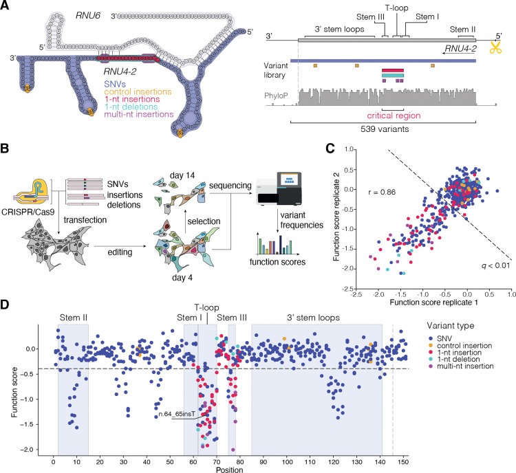
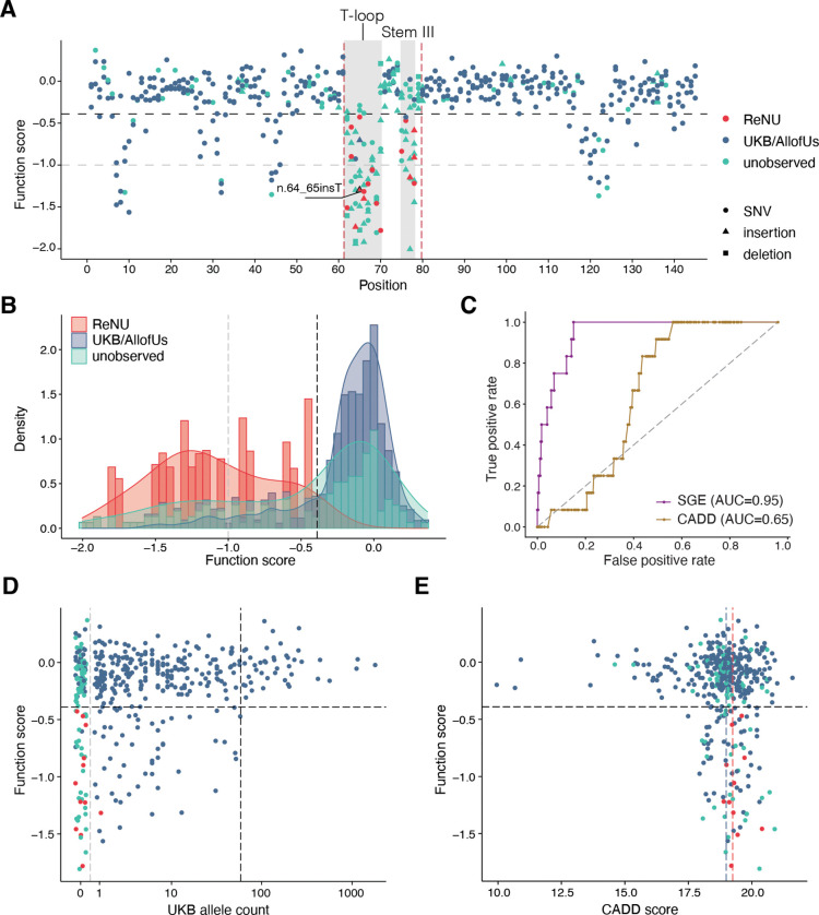
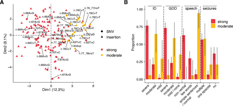
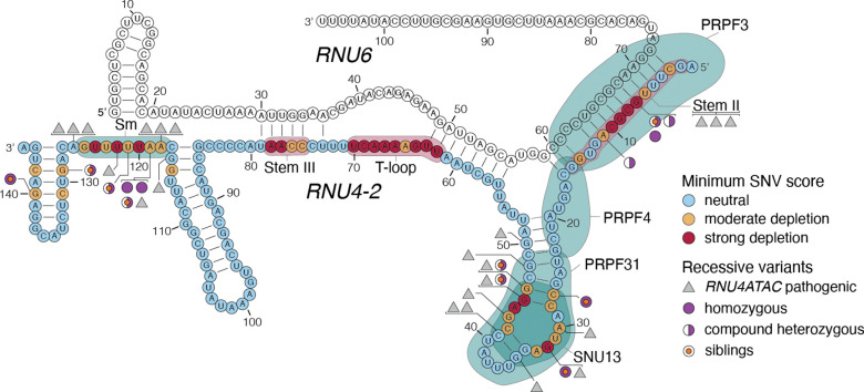
Recently, de novo variants in an 18 nucleotide region in the centre of RNU4-2 were shown to cause ReNU syndrome, a syndromic neurodevelopmental disorder (NDD) that is predicted to affect tens of thousands of individuals worldwide1,2. RNU4-2 is a non-protein-coding gene that is transcribed into the U4 small nuclear RNA (snRNA) component of the major spliceosome3. ReNU syndrome variants disrupt spliceosome function and alter 5' splice site selection1,4. Here, we performed saturation genome editing (SGE) of RNU4-2 to identify the functional and clinical impact of variants across the entire gene. The resulting SGE function scores, derived from variants' effects on cell fitness, discriminate ReNU syndrome variants from those observed in the population and dramatically outperform in silico variant effect prediction. Using these data, we redefine the ReNU syndrome critical region at single nucleotide resolution, resolve variant pathogenicity for variants of uncertain significance, and show that SGE function scores delineate variants by phenotypic severity. Further, we identify variants impacting function in regions of RNU4-2 that are critical for interactions with other spliceosome components. We show that these variants cause a novel recessive NDD that is clinically distinct from ReNU syndrome. Together, this work defines the landscape of variant function across RNU4-2, providing critical insights for both diagnosis and therapeutic development.

Keywords: ReNU syndrome; Saturation genome editing; clinical variant interpretation; neurodevelopmental disorders; non-coding RNA; recessive; small nuclear RNA.


Figures

Figure 1.
Figure 1.. Saturation Genome Editing reveals the functional spectrum of RNU4–2 variants.
A. Schematic of SGE library design and CRISPR targeting strategy for RNU4–2. Positions of library variants including all possible SNVs (navy; across the 145-nt transcript and 6-nt 3’), control 1-nt insertions in loop regions (yellow), critical region (CR) 1-nt insertions (red) and deletions (teal) and multi-nt insertions (purple) are denoted on a schematic of RNU4–2 and RNU6 in complex (left) and by genomic location (right). A gRNA was designed to cleave upstream of RNU4–2 (scissors), avoiding highly repetitive sequence and allowing for a PAM-blocking variant to be installed in a region of low conservation. (PhyloP 100 vertebrates basewise conservation track shown.) B. Schematic of SGE experiments in HAP1. Following editing, cells were harvested on days 4 and 14. Sequencing was performed to quantify variant frequencies at each timepoint and function scores were calculated. C. Function scores for 539 variants were correlated across biological replicates (Pearson’s r = 0.86). Variants scoring significantly lower than control insertions (q < 0.01) are indicated with the dashed line. D. Function scores are plotted by genomic position in relation to RNU4–2 (RefSeq: NR_003137.3). The line at n.145 marks the end of the transcript, with 18 more distal SNVs also scored.
Figure 2.
Figure 2.. Function scores accurately discriminate variants underlying ReNU syndrome.
A. Function scores for 521 variants within the RNU4–2 transcript are plotted by position and coloured by their association with ReNU syndrome (red), presence in the UK Biobank or All of Us cohorts (blue), or no observation in either (teal). Depleted variants within the 18-nt CR (marked by vertical red dashed lines) are confined to two smaller regions (shaded grey) and include all ReNU syndrome variants scored (n = 18). These regions, n.62–70 and n.75–78, correspond to the T-loop and Stem III, respectively. The black dashed line (function score = −0.39) indicates significantly depleted variants and the gray dashed line (function score = −1.00) separates “moderate” from “strong” depletion. B. Stacked histogram and overlaid density plot of function scores by category comparing 18 ReNU syndrome variants to 348 variants observed in UK Biobank and/or All of Us and 155 unobserved variants. C. ROC curves show the performance of SGE function scores and CADD scores for classifying ReNU syndrome SNVs (n = 12) from SNVs observed at least once in population controls (n = 346). D. Function scores for SNVs are plotted by UK Biobank allele count (AC). Higher allele counts were correlated with higher function scores (Spearman’s ρ = 0.19, P = 5.3 × 10⁻⁵). Among 43 SNVs with allele count > 59 (black dashed line), no SNVs were depleted. The gray dashed line separates variants absent from UK Biobank (AC = 0) from those observed (AC > 0). E. Function scores for 435 SNVs are plotted by CADD score. The dashed line at y = −0.39 indicates significantly depleted SNVs, whereas the red line at x = 19.25 and the blue line at x = 18.99 indicate median CADD scores for ReNU syndrome SNVs and SNVs present in population cohorts, respectively.
Figure 3.
Figure 3.. Function scores predict ReNU syndrome severity.
A. The first two principal components from clustering of 143 ReNU syndrome cases by phenotype using the approach from Nava et al. Variants are coloured by SGE function score class (strong depletion: function score < −1.0, moderate depletion: −1 < function score < −0.39). Unlabelled triangles indicate occurrences of n.64_65insT. B. The proportion of affected individuals with each phenotype is plotted, with cases grouped by SGE function score class. Error bars indicate 95% confidence intervals. ID: intellectual disability; GDD: global developmental delay.
Figure 4.
Figure 4.. SGE-depleted variants outside the CR cause a recessive NDD.
The lowest SGE function score class among SNVs at each position is indicated on the U4:U6 secondary structure. Outside the CR, low SGE scores occur at positions of spliceosomal protein binding, indicated by teal shaded regions. Black triangles correspond to homologous positions of RNU4ATAC at which (likely) pathogenic variants have been linked to recessive disease (from ClinVar; Sup. Table 5). RNU4–2 variants with low function scores observed in recessive NDD cases are indicated, with filled purple circles indicating variants observed as homozygous and half-filled circles indicating variants observed in the compound heterozygous state. An orange dot in the centre of a circle indicates that the variant is observed in two affected siblings. Six (likely) pathogenic RNU4ATAC variants could not be confidently assigned to an equivalent nucleotide in RNU4–2. Three of these (n.8C>A, n.13C>T, and n.16G>A) are shown together as mapping to Stem II. The other three (n.29T>G, n.30G>A, and n.111G>A) are not shown. U4/U6 structure depiction adapted from Quinodoz et al.

References

    1. Chen Y. et al. De novo variants in the RNU4–2 snRNA cause a frequent neurodevelopmental syndrome. Nature (2024) doi: 10.1038/s41586-024-07773-7. - DOI - PMC - PubMed
    1. Greene D. et al. Mutations in the U4 snRNA gene RNU4–2 cause one of the most prevalent monogenic neurodevelopmental disorders. Nat. Med. (2024) doi: 10.1038/s41591-024-03085-5. - DOI - PMC - PubMed
    1. Nguyen T. H. D. et al. The architecture of the spliceosomal U4/U6.U5 tri-snRNP. Nature 523, 47–52 (2015). - PMC - PubMed
    1. Nava C. et al. Dominant variants in major spliceosome U4 and U5 small nuclear RNA genes cause neurodevelopmental disorders through splicing disruption. medRxiv 2024.10.07.24314689 (2024) doi: 10.1101/2024.10.07.24314689. - DOI - PMC - PubMed
    1. Bruselles A. et al. Expanding the mutational spectrum of ReNU syndrome: insights into 5’ Stem-loop variants. Eur. J. Hum. Genet. 1–9 (2025). - PMC - PubMed

Publication types

LinkOut - more resources