. 2025 Apr 14:16:1569490.
doi: 10.3389/fimmu.2025.1569490. eCollection 2025.

Multiplex immunofluorescence assessment of macrophages and IL-23R in inflammatory and malignant diseases of the oral mucosa: a pilot study

Affiliations

Multiplex immunofluorescence assessment of macrophages and IL-23R in inflammatory and malignant diseases of the oral mucosa: a pilot study

Leah Trumet et al. Front Immunol. .

Abstract

Background: Immune cells play a major role in the development and progression of inflammatory and malignant diseases of the oral mucosa. There is growing evidence that immune cells contribute to oral cancer progression and metastases. Inflammatory carcinogenesis is believed to be relevant for oral Lichen Planus as well as for oral Leukoplakia. In addition, there is growing evidence that periodontitis might also be linked to oral cancer development. Yet there is no analysis available comparing the immune cell composition in these different inflammatory and malignant neoplastic diseases. A better understanding of similarities and differences of the diseases could eventually also pave the way for the use of immunotherapy in non-malignant diseases.

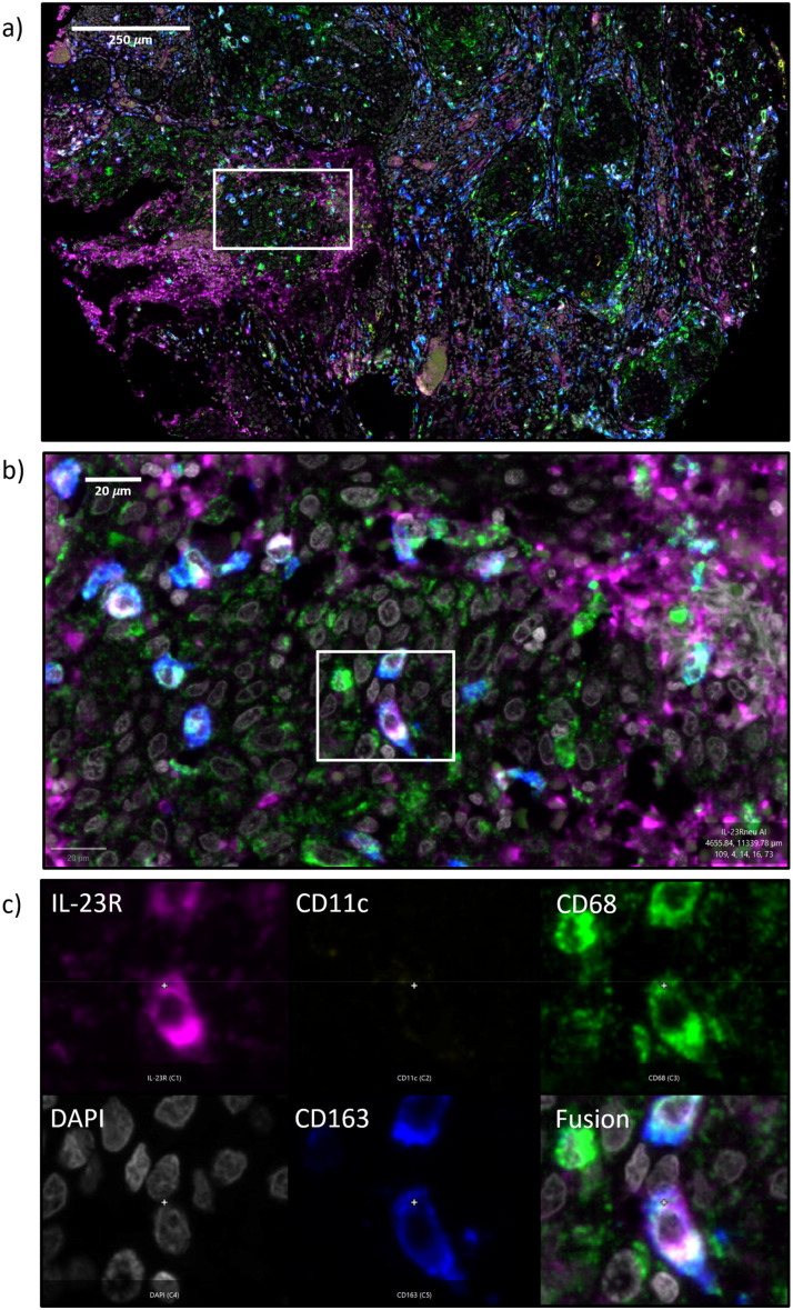
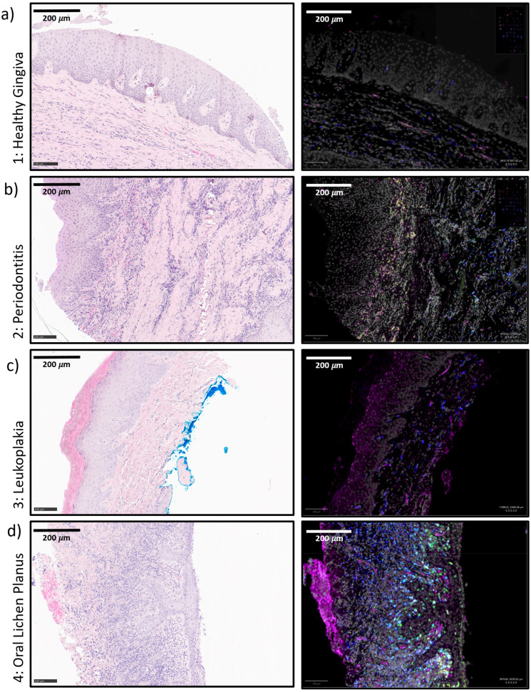
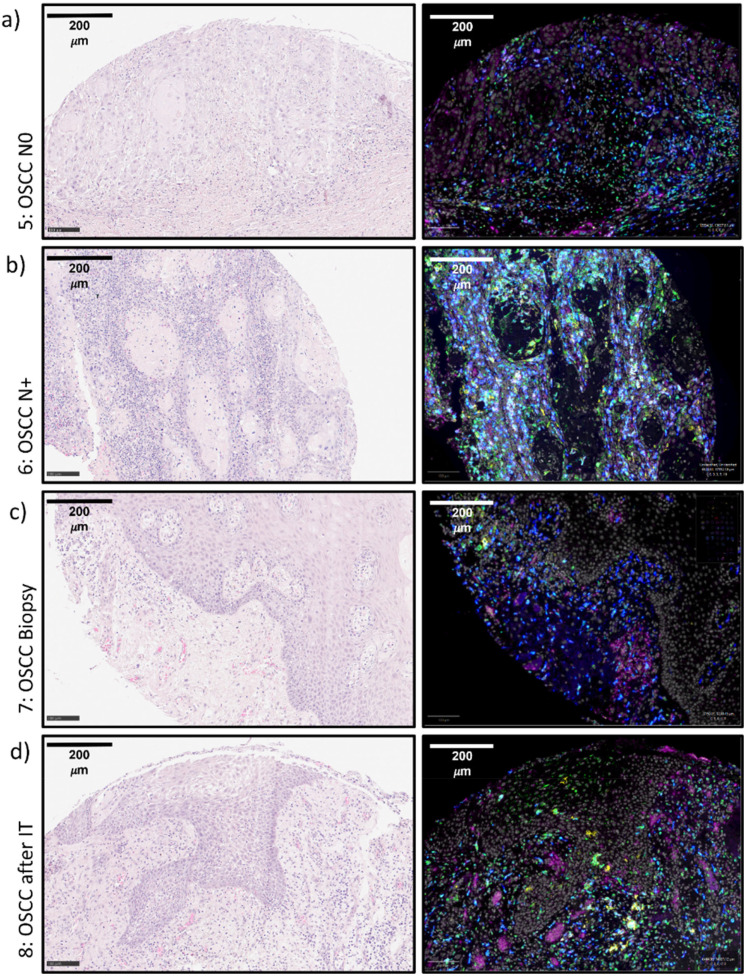
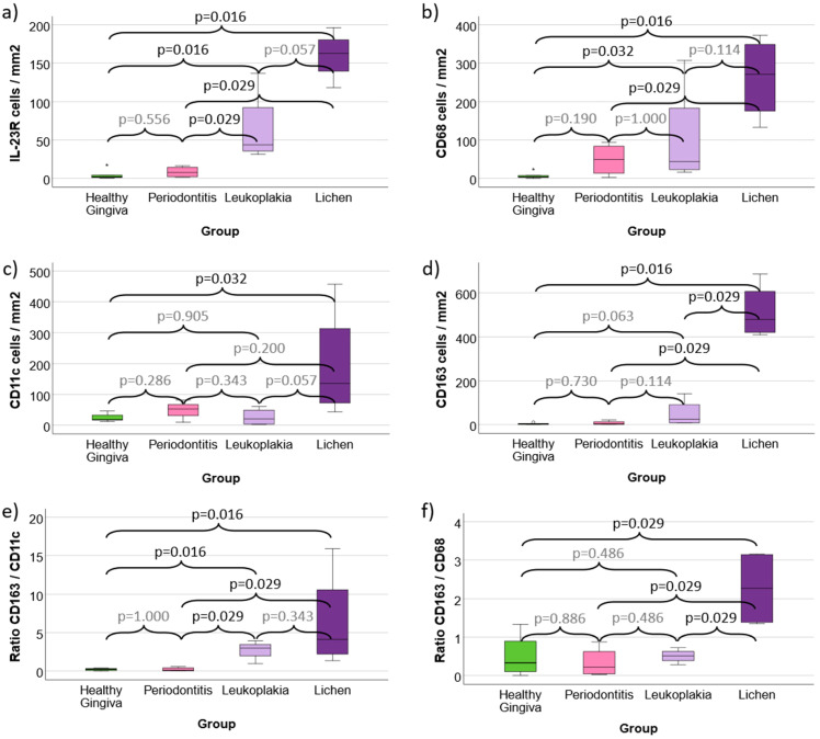
Methods: In the current pilot study, a tissue microarray (TMA) was created of a total of 29 patients with periodontitis (PD, n=4), oral Leukoplakia (OL, n=4), oral Lichen Planus (OLP, n=4), oral squamous cell cancer without lymphatic metastases (OSCC N0, n=5), or with lymphatic metastases (OSCC N+, n=4), OSCC biopsies prior to and resection specimens after anti-PD1 immunotherapy (IT) (each n=3) as well as healthy control gingiva (n=5). In each patient two tissue samples were analyzed. The TMA was stained with a 4X multiplex immunofluorescent staining for IL-23R, CD68, CD11c, and CD163. Samples were digitalized and an AI-based cell counting was performed. Statistical analysis was performed using the Mann-Whitney U test.

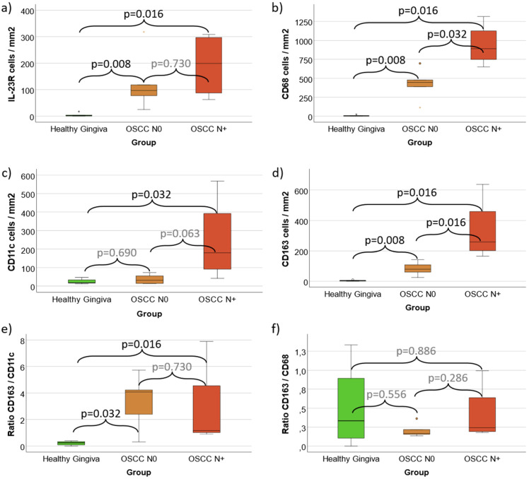
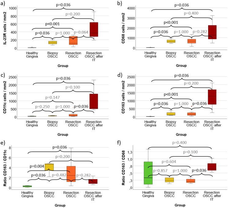
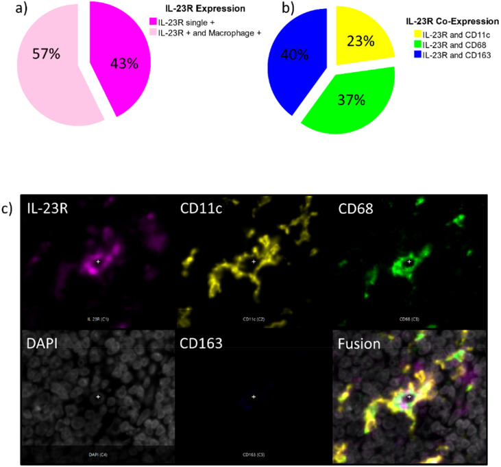
Results: IL-23R expression, macrophage infiltration as well as M2 polarization in OL and OLP were significantly higher compared to controls. OLP showed a significantly higher M2 infiltration and polarization than OL. PD showed a trend for increased macrophage infiltration compared to controls without significance. N+ OSCC showed a significantly increased macrophage infiltration compared to N0 cases. In response to anti-PD1 IT, CD11c and CD163 infiltration was significantly increased. Most IL-23R positive cells co-expressed macrophage markers.

Conclusion: A TMA in combination with 4-plex immunofluorescence is suitable for immune cell characterization in different oral diseases. Macrophage infiltration and polarization in precursor lesions seems to be associated with OSCC development as well as metastatic spread. IL-23 pathway inhibition might be a potential target for oral Lichen and Leukoplakia.

Keywords: Lichen Planus; N+; OSCC; leukoplakia; lymph node metastases; oral cancer; oral medicine; periodontitis.


Conflict of interest statement

The authors declare that the research was conducted in the absence of any commercial or financial relationships that could be construed as a potential conflict of interest. The author(s) declared that they were an editorial board member of Frontiers, at the time of submission. This had no impact on the peer review process and the final decision.

Figures

Figure 1
Figure 1
(a–c) Immunofluorescence 4x multi-staining The figure shows an exemplary micrograph of an oral squamous cell carcinoma (OSCC) tumor resection specimen ( Figure 1a ) and a high-power magnification (Fib. 1B). Figure 1c shows a magnification on single-cell level (200x magnification) and a separate image for each fluorescence channel: IL-23R (magenta), CD11c (yellow), CD68 (green), DAPI (white), CD163 (blue) and a fusion image.
Figure 2
Figure 2
(a–d) Exemplary tissue micrographs (1-4) This figure shows exemplary micrographs for each analyzed tissue. On the left column, HE-stainings are displayed. On the right column, the corresponding immunofluorescence stainings are given. The immunofluorescence fusion images show DAPI (white), IL-23R (magenta), CD11c (yellow), CD68 (green) and CD163 (blue). (a) 1: Healthy Control Gingiva (b) 2: Periodontitis (c) 3: Oral Leukoplakia (d) 4: Oral Lichen Planus.
Figure 3
Figure 3
(a–d) Exemplary tissue micrographs (5-8) This figure shows exemplary micrographs for each analyzed tissue. On the left column, HE-stainings are displayed. On the right column, the corresponding immunofluorescence stainings are given. The immunofluorescence fusion images show DAPI (white), IL-23R (magenta), CD11c (yellow), CD68 (green) and CD163 (blue). (a) 5: OSCC N0 (b) 6: OSCC N+ (c) 7: OSCC Biopsy (d) 8: OSCC Resection after IT OSCC, oral squamous cell carcinoma; IT, Immunotherapy with anti-PD1 antibodies.
Figure 4
Figure 4
(a–f) Cell density and expression ratios in Periodontitis, oral Leukoplakia, oral Lichen Planus and healthy Gingiva controls (a) Cell density of IL-23 Receptor expressing cells (b) Cell density of CD68 positive macrophages (c) Cell density of CD11c positive macrophages (predominantly M1-like) and dendritic cells (d) Cell density of CD163 positive macrophages (predominantly M2-like) (e) CD163 / CD11c expression ratio as indication of M2 polarization (f) CD163 / CD68 expression ratio as indication of M2 polarization Values for Healthy Gingiva, Periodontitis, oral Leukoplakia and oral Lichen Planus are given. Figures 4a-d shows the cell density (cells/mm2 specimen area) of IL-23R, CD68, CD11c and CD163 positive cells. Figures 4e, f give the rations of all CD163 expressing cells versus all CD11c and CD68 expressing cells as indicator of M2 polarization of macrophages in the analyzed tissues. p-values generated using the Mann-Whitney U test are shown.
Figure 5
Figure 5
(a–f) Cell density and expression ratios in N0 OSCC and N+ OSCC compared with controls (a) Cell density of IL-23 Receptor expressing cells (b) Cell density of CD68 positive macrophages (c) Cell density of CD11c positive macrophages (predominantly M1-like) and dendritic cells (d) Cell density of CD163 positive macrophages (predominantly M2-like) (e) CD163 / CD11c expression ratio as indication of M2 polarization (f) CD163 / CD68 expression ratio as indication of M2 polarization Values for Healthy Gingiva, oral squamous cell carcinoma (OSCC) resection specimens without lymph node metastases (N0) and cases with lymph node metastases (N+) are given. Figures 5a-d shows the cell density (cells/mm2 specimen area) of IL-23R, CD68, CD11c and CD163 positive cells. Figures 5e, f give the rations of all CD163 expressing cells versus all CD11c and CD68 expressing cells as indicator of M2 polarization of macrophages in the analyzed tissues. p-values generated using the Mann-Whitney U test are shown.
Figure 6
Figure 6
(a–f) Cell density and expression ratios in OSCC Biopsies, Resection specimens and Resection specimens after anti-PD1 immunotherapy (IT) compared with controls (a) Cell density of IL-23 Receptor expressing cells (b) Cell density of CD68 positive macrophages (c) Cell density of CD11c positive macrophages (predominantly M1-like) and dendritic cells (d) Cell density of CD163 positive macrophages (predominantly M2-like) (e) CD163 / CD11c expression ratio as indication of M2 polarization (f) CD163 / CD68 expression ratio as indication of M2 polarization Values for Healthy Gingiva, oral squamous cell carcinoma (OSCC) Biopsy specimens, OSCC tumor resection samples (irrespective of the N-Status) and tumor resection specimens after anti-PD1 immunotherapy (IT) are given. Biopsy samples and specimens after IT were obtained from the same patients. Figures 6a-d shows the cell density (cells/mm2 specimen area) of IL-23R, CD68, CD11c and CD163 positive cells. Figures 6e, f give the rations of all CD163 expressing cells versus all CD11c and CD68 expressing cells as indicator of M2 polarization of macrophages in the analyzed tissues. p-values generated using the Mann-Whitney U test are shown.
Figure 7
Figure 7
(a–c) Lineage of IL-23R expressing cells Figure 7a shows the percentage of IL-23R single positive cells compared to the Il-23R positive cells that co-express macrophage and Dendritic Cell markers (CD68, CD163, CD11c) in all analyzed tissue specimens. Figure 7b differentiates the IL-23 co-expressing cells in IL-23R and CD11c positive cells (yellow), IL-23R and CD68 positive cells (blue) as well as IL-23R and CD163 positive cells (blue). Figure 7c shows an exemplary magnification on single-cell level (200x) showing a CD11c+ CD68+ IL-23+ Dendritic Cell with typical morphology. A separate image for each fluorescence channel IL-23R (magenta), CD11c (yellow), CD68 (green), DAPI (white), CD163 (blue) and a fusion image are given.

Similar articles

References

    1. Sun Y, Liu N, Guan X, Wu H, Sun Z, Zeng H. Immunosuppression induced by chronic inflammation and the progression to oral squamous cell carcinoma. Mediators Inflammation. (2016) 2016:5715719. doi: 10.1155/2016/5715719 - DOI - PMC - PubMed
    1. Baima G, Minoli M, Michaud DS, Aimetti M, Sanz M, Loos BG, Romandini M, et al. . Periodontitis and risk of cancer: Mechanistic evidence. Periodontol 2000. (2024) 96(1):83–94. doi: 10.1111/prd.12540 - DOI - PMC - PubMed
    1. Li C, Dong X, Li B. Tumor microenvironment in oral squamous cell carcinoma. Front Immunol. (2024) 15:1485174. doi: 10.3389/fimmu.2024.1485174 - DOI - PMC - PubMed
    1. Luo M, Zhao F, Cheng H, Su M, Wang Y. Macrophage polarization: an important role in inflammatory diseases. Front Immunol. (2024) 15:1352946. doi: 10.3389/fimmu.2024.1352946 - DOI - PMC - PubMed
    1. Wang Y, Xu B, Hu WW, Chen LJ, Wu CP, Lu BF, et al. . High expression of CD11c indicates favorable prognosis in patients with gastric cancer. World J Gastroenterol. (2015) 21(31):9403–12. doi: 10.3748/wjg.v21.i31.9403 - DOI - PMC - PubMed

MeSH terms

Substances