. 2025 May;97(5):e70372.
doi: 10.1002/jmv.70372.

Outbreak of Enterovirus D68 in Young Children, Brescia, Italy, August to November 2024

Affiliations

Outbreak of Enterovirus D68 in Young Children, Brescia, Italy, August to November 2024

Serena Messali et al. J Med Virol. 2025 May.

Abstract

Enterovirus D68 (EV-D68) is responsible for a plethora of clinical manifestations ranging from asymptomatic infections to severe respiratory symptoms and neurological disorders. EV-D68 was first detected in children with pneumonia in 1962 and, from then, only sporadic cases were reported until 2014, when outbreaks were notified across the world. After the withdrawal of preventive measures against SARS-CoV-2, a significant increase in EV-D68 infections has been reported in 2021-2022. A surveillance program to evaluate the incidence of enterovirus/rhinovirus (EV/RV) infections was implemented at the Brescia Civic Hospital, Italy. Fifty-five EV/RV-positive respiratory samples, belonging to pediatric patients, were subjected to NGS. We observed that 61.8% of samples were positive for EV, with EV-D68 as the most prevalent genotype predominantly detected between August and November 2024. Phylogenetic analysis revealed that EV-D68 sequences formed two monophyletic clades corresponding to the A2 and B3 lineages, highlighting their recent introduction in Italy. Interestingly, 40% of pediatric EV-D68 infections were detected with at least one other EV/RV. Our study highlights the crucial role played by genomic surveillance of respiratory infections to monitor the circulation of emerging and re-emerging viruses, as well as their evolution. This will be fundamental to enable prompt intervention strategies.

Keywords: children; clinical presentation; enterovirus D68; epidemiology; phylogenetic analysis.


Conflict of interest statement

The authors declare no conflicts of interest.

Figures
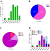
Figure 1
Figure 1
Distribution and frequency of EV/RV from May to December 2024 in the Brescia area. (A) The graph shows the spread of EV/RV‐positive cases in the pediatric population. (B) Pie chart displays the frequency of EV species as compared to RV ones. (C) Pie chart illustrates the percentage of each EV species. (D) The graph shows the monthly distribution, from May to December 2024, of EV/RV genotypes. RV genotypes are grouped according to their corresponding species (A–C). CV, coxsackievirus; E, echovirus; EV, enterovirus; RV, rhinovirus.
Figure 2
Figure 2
Phylogenetic relationships and temporal dynamics of EV‐D68. (A) Maximum likelihood (ML) phylogenetic tree of EV‐D68 genomes (n = 1316), including 25 novel Brescia isolates. Lineages are color‐coded. The heatmap surrounding the tree represents the geographic origin of the samples: Africa (brown), Asia (green), Europe (yellow), Italy (red), North America (gray), and Oceania (light blue). The outer ring indicates the sampling dates, ranging from 2000 to 2024. Two well‐supported monophyletic clades were identified: Clade I (A2) and Clade II (B3). (B) Bayesian MCC tree for Clade I (A2 lineage), showing Brescia isolates clustering with recent sequences from Europe, Asia, and North America. Posterior support values are indicated at key nodes. (C) Maximum clade credibility (MCC) tree for Clade II (B3 lineage), displaying a well‐supported cluster of Brescia isolates closely related to North American sequences. The shaded density plot represents the posterior distribution of the most recent common ancestor.
Figure 3
Figure 3
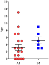
Age distribution of EV‐D68 A2 and B3 lineages. The graph represents the distribution of EV‐D68‐positive samples according to patients’ age for each lineage.

References

    1. Grizer C. S., Messacar K., and Mattapallil J. J., “Enterovirus‐D68 ‐ A Reemerging Non‐Polio Enterovirus That Causes Severe Respiratory and Neurological Disease in Children,” Frontiers in Virology 4 (2024): 1328457, 10.3389/fviro.2024.1328457. - DOI - PMC - PubMed
    1. Elrick M. J., Pekosz A., and Duggal P., “Enterovirus D68 Molecular and Cellular Biology and Pathogenesis,” Journal of Biological Chemistry 296 (2021): 100317, 10.1016/j.jbc.2021.100317. - DOI - PMC - PubMed
    1. Midgley S. E., Benschop K., Dyrdak R., et al., “Co‐Circulation of Multiple Enterovirus D68 Subclades, Including a Novel B3 Cluster, Across Europe in a Season of Expected Low Prevalence, 2019/20,” Eurosurveillance 25, no. 2 (2020): 1900749, 10.2807/1560-7917.ES.2020.25.2.1900749. - DOI - PMC - PubMed
    1. Schieble J. H., Fox V. L., and Lennette E. H., “A Probable New Human Picornavirus Associated With Respiratory Disease1,” American Journal of Epidemiology 85, no. 2 (1967): 297–310, 10.1093/oxfordjournals.aje.a120693. - DOI - PubMed
    1. Holm‐Hansen C. C., Midgley S. E., and Fischer T. K., “Global Emergence of Enterovirus D68: A Systematic Review,” Lancet Infectious Diseases 16, no. 5 (2016): e64–e75, 10.1016/S1473-3099(15)00543-5. - DOI - PubMed

LinkOut - more resources