. 2025 Jun 11;16(6):e0098425.
doi: 10.1128/mbio.00984-25. Epub 2025 Apr 29.

Role of glucuronoxylomannan and steryl glucosides in protecting against cryptococcosis

Affiliations

Role of glucuronoxylomannan and steryl glucosides in protecting against cryptococcosis

Gabriel Soares Matos et al. mBio. .

Abstract

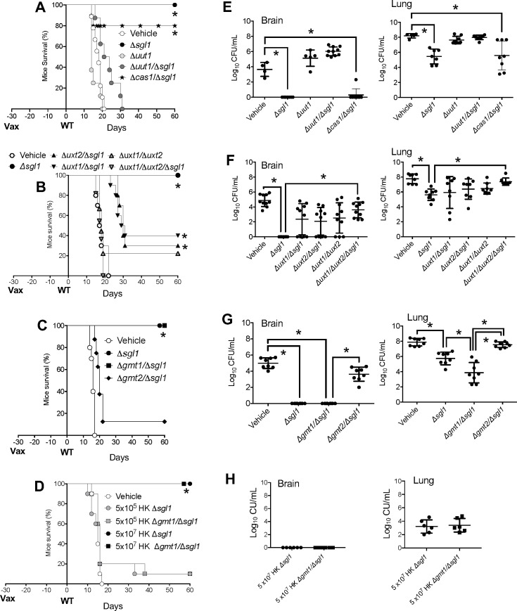
The development of vaccines for fungal diseases, including cryptococcosis, is an emergent line of research and development. In previous studies, we showed that a Cryptococcus mutant lacking the SGL1 gene (∆sgl1) accumulates certain glycolipids called steryl glucosides (SGs) on the fungal capsule, promoting an effective immunostimulation that totally protects the host from a secondary cryptococcal infection. However, this protection is lost when the cryptococcal capsule is absent in the ∆sgl1 background. The cryptococcal capsule is mainly composed of glucuronoxylomannan (GXM), a polysaccharide microfiber consisting of glucuronic acid, xylose, and mannose linked by glycosidic bonds forming specific triads. In this study, we engineered cells to lack each of the GXM components and tested the effect of these deletions on protection under the condition of SG accumulation. We found that glucuronic acid and xylose are required for protection, and their absence abrogates the production of IFNγ and IL-17A by γδ T cells, which are necessary stimulants for the protective phenotype of the ∆sgl1. We analyzed the structure of the GXM microfibers and found that although the deletion of SGL1 only slightly affects the size and distribution of these microfibers, it significantly changes the ratio of mannose to other components. In conclusion, this study identifies the structural modifications that the deletion of SGL1 and the consequent accumulation of SGs impart to the GXM structure of C. neoformans. This provides significant insights into the protective mechanisms mediated by SG accumulation on the capsule, with important implications for the future development of an efficacious cryptococcal vaccine.IMPORTANCECryptococcus neoformans is an encapsulated fungus that causes invasive fungal infections with high morbidity and mortality in susceptible patients. With increasing drug resistance and high toxicity of current antifungal drugs, there is a need for alternative therapeutic strategies, such as a cryptococcal vaccine. In this study, we identify the necessary capsular components and their structural organization required for a cryptococcal vaccine to protect the host against challenge with a virulent strain. These capsular components are glucuronic acid, xylose, and mannose, and they work together with certain glycolipids called steryl glucosides (SGs) to stimulate host immunity. Interestingly, SGs on the capsule may favor the formation of small capsular microfibers organized in specific mannose triads. Thus, the results of this paper are important because they identify a mechanism by which SGs affect the structure of the cryptococcal capsule, with important implications for the future development of a cryptococcal vaccine using capsular components and SGs.

Keywords: Cryptococcus neoformans; fungal infection; glucuronic acid; glucuronoxylomannan; immunity; mannose; steryl glucosides; vaccine; xylose.


Conflict of interest statement

Maurizio Del Poeta is a co-founder and the chief scientific officer of MicroRid Technologies Inc. All other authors declare no competing interests.

Figures

Fig 1
Fig 1
Diagram of the polysaccharide capsule of C. neoformans. GXM is made by sugar polymerization into an elongated carbohydrate backbone, from UDP-glucuronic acid, UDP-xylose, and GDP-mannose precursors. Precursor synthesis occurs in the cytoplasm. As shown at the lower right, GDP-mannose is produced through the activity of Man1, a phosphomannose isomerase. UDP-glucose is converted to UDP-glucuronic acid by UDP-glucose dehydrogenase (Ugd1) and then to UDP-xylose by Uxs1 decarboxylase. UDP-glucose can also be converted to UDP-galactose, which is required for GXMGal synthesis, in a reaction catalyzed by Uge1 epimerase. As shown at the bottom left, nucleotide sugars are then transported into the Golgi by Gmt1 and Gmt2 (GDP-mannose), Uxt1 and Uxt2 (UDP-xylose), and Uut1 (UDP-glucuronic acid). Mannosylation is performed by an α−1,3 mannosyltransferase (1), and the formation of a β−1,2 glycosidic bond between glucuronic acid and mannose is catalyzed by β−1,2 glucuronyltransferase (2); xylosyl can be added by β−1,2 and/or β−1,4 xylosyltransferase activities (3). O-acetyl residues are also added to the growing polysaccharide by Cas1. This sequence of events is putative. The number of xylose side chains and the amount of 6-O-acetylation varies with strain and growth conditions. After synthesis, GXM molecules exit the cell and associate with the cell wall.
Fig 2
Fig 2
Effect on capsule size when SGL1 is deleted from strains lacking transporters of GXM precursors. Strains were grown in the capsule-inducing medium at pH 7.4, and capsule measurements were performed by India ink staining. (A) Samples were imaged using a Zeiss microscope, and pictures were taken using Zen Pro software. (B) Capsule thickness was determined as described in Materials and Methods. *, P < 0.05, versus wild type (WT) or ∆sgl1; %, P < 0.05 between the indicated strains. Significance was determined via a one-way ANOVA using Dunnett’s multiple comparison tests for P value adjustment.
Fig 3
Fig 3
Transport of GXM components affects SG localization. Ergosterol-3β-D-glucoside (the main component of SG), extracted from cell pellet (A), extracellular medium (B), and GXM fraction (C) was analyzed by liquid chromatography mass spectrometry (LC-MS). Uut1, Uxt1, Uxt2, and Gmt2 are required for SGs to be associated with the GXM fraction. *, P < 0.05, versus wild-type WT) strain; #, P < 0.05 versus ∆sgl1 strain. Significance was determined via one-way ANOVA using Dunnett’s multiple comparison tests for P value adjustment.
Fig 4
Fig 4
Deletion of the SGL1 gene renders the respective double mutant avirulent. Mice survival (A-D) and tissue burden culture (TBC) by colony forming unit (CFU) analysis (E-H) of CBA/J mice infected intranasally with 5 × 10⁵ C. neoformans cells. The ∆uut1/sgl1,uxt1/sgl1,uxt2/sgl1,gmt1/sgl1,gmt2/sgl1, and ∆cas1/sgl1 double mutants are avirulent, like the ∆sgl1 mutant. Average survival is as follows: (A) WT 16.7 ± 1.49 days; (B) WT 16 ± 2 days, ∆uxt1 16 ± 1.56 days, ∆uxt2 15.2 ± 2.74 days; (C) WT 16.9 ± 1.1 days, ∆gmt1 17 ± 1.82 days, ∆gmt2 17.9 ± 2.02 days; (D) WT 14.6 ± 1.17 days, ∆cas1 19.2 ± 2.97 days. In A, B, C, and D *, P < 0.05 versus wild type (WT) strain. Statistical analysis performed by rank (Mantel-Cox) test. Values are expressed as mean ± standard error. In E, F, G, and H, *, P < 0.05 between the indicated strains. Values are expressed as mean ± standard error. Statistical analysis was conducted using one-way analysis of variance (ANOVA), followed by Tukey’s post hoc test for multiple comparisons.
Fig 5
Fig 5
Deletion of SGL1 abrogates cryptococcal virulence. CBA/J mice were intranasally infected with 5 × 10⁵ C. neoformans cells, and at the indicated days, three mice per strain were euthanized, and their organs were collected and processed for tissue burden analysis using CFUs. CFU analysis was performed at 3, 6, 9, 14, 21, and 30 days post-infection for lungs (A-D) and brain (E). Values are expressed as mean ± standard error. *, P < 0.05 between the indicated strain versus the WT (in A) or versus ∆sgl1 (in B-D). In E, *, P < 0.05 between the WT versus any mutant. Statistical analysis was conducted using one-way analysis of variance (ANOVA), followed by Tukey’s post hoc test for multiple comparisons.
Fig 6
Fig 6
Deletion of GXM transporters affects vaccine protection when SGs accumulate. Survival (A-D) and CFU analysis (E-H) of CBA/J mice vaccinated (Vax) with 5 × 10⁵ cells of avirulent C. neoformans mutant(s) and challenged after 30 days with 5 × 10⁵ cells of wild-type (WT) C. neoformans. Only ∆sgl1 and ∆gmt1/sgl1 administration provided full protection. Average survival is as follows: (A) vehicle 18 ± 2.44 days, ∆uut1 15.8 ± 2.69 days, and ∆uut1/sgl1 23 ± 5.41 days; (B) vehicle 18.2 ± 2.82 days, and ∆uxt1/uxt2/sgl1 17.5 ± 1.5 days; (C) vehicle 17.6 ± 1.64 days; (D) vehicle 17.1 ± 1.19 days. In A, B, C, and D *, P < 0.05 versus wild-type (WT) strain. Statistical analysis performed by rank (Mantel-Cox) test. Values are expressed as mean ± standard error. In E, F, G, and H, *, P < 0.05 between the indicated strains. Values are expressed as mean ± standard error. Statistical analysis was conducted using one-way analysis of variance (ANOVA), followed by Tukey’s post hoc test for multiple comparisons.
Fig 7
Fig 7
γδ T cell response is affected by the deletion of nucleotide sugar transporter. Evaluation of inflammatory interleukin secretion by γδ T cells. IFNγ (A) and IL-17A (B) were measured after a 5-day exposure of γδ T cells to capsule mutants. Glucuronic acid, xylose, and mannose organization on the cryptococcal capsule is important to stimulate a protective cytokine response by γδ T cells in the presence of SGs. *P < 0.05 versus the WT strain; #, P < 0.05 versus the ∆sgl1 strain; $, P < 0.05 versus the indicated groups; % P < 0.05 versus WT; ns, not significant. Values are expressed as mean ± standard error. Statistical analysis was conducted using one-way analysis of variance (ANOVA), followed by Tukey’s post hoc test for multiple comparisons.
Fig 8
Fig 8
Analysis of mAb 18B7 staining of C. neoformans capsular surface. C. neoformans was grown in capsule-inducing conditions and stained with anti-GXM antibody 18B7 followed by a fluorescent secondary antibody conjugated to Alexa Fluor 568 (red). Cell wall chitin was stained using calcofluor white (in blue). (A) Images were taken at 100× magnification using a Zeiss Axio observer microscope (Thornwood, NY). (B) Fluorescence intensity was quantified, and the average fluorescence was measured using ImageJ/Fiji software. Mean fluorescence intensity values (MFI) derived from at least 50 cells were normalized by subtracting the MFI of the acapsular strain ∆cap59, which was the control for the capsular staining background. Results show that SG accumulation affects the binding of 18B7 to GXM when the level of glucuronic acid, xylose, or mannose is altered. *, P < 0.05 versus WT. Values are expressed as mean ± standard error. Statistical analysis was conducted using one-way ANOVA, followed by Tukey’s post hoc test for multiple comparisons.
Fig 9
Fig 9
Size distribution of capsular GXM by DLS. The size distribution of GXM from C. neoformans wild-type (WT), ∆sgl1, and nucleotide-sugar transporter mutants ∆gmt1, ∆gmt1/∆sgl1, ∆gmt2, and ∆gmt2/∆sgl1 was analyzed using DLS. The x-axis represents the measured particle size distribution, whereas the y-axis corresponds to the intensity-weighted size percentages. Results suggest that the accumulation of SGs may affect the peak shape without causing a major difference in GXM size. The results shown are representative of at least three separate experiments.
Fig 10
Fig 10
Accumulation of SGs alters GXM architecture by 1D-NMR spectra analysis. (A) Comparison ofα-mannose anomeric region of 1D-NMR spectra of the GXM isolated from C. neoformans WT, ∆sgl1, ∆gmt1/∆sgl1, and ∆gmt2/∆sgl1. Red arrows indicate the absence of M1 in ∆sgl1 and ∆gmt1/∆sgl1 strains. The spectra indicate that the ∆sgl1 and the ∆gmt1/∆sgl1 mutant lack the M1 mannose triad, which is the major triad in the WT strain. (B) Composition of the four types of mannose triads. (C) Measurement of the mol% of the different mannose triad elements estimated by 1D-NMR analysis.

References

    1. Rajasingham R, Smith RM, Park BJ, Jarvis JN, Govender NP, Chiller TM, Denning DW, Loyse A, Boulware DR. 2017. Global burden of disease of HIV-associated cryptococcal meningitis: an updated analysis. Lancet Infect Dis 17:873–881. doi: 10.1016/S1473-3099(17)30243-8 - DOI - PMC - PubMed
    1. Stott KE, Loyse A, Jarvis JN, Alufandika M, Harrison TS, Mwandumba HC, Day JN, Lalloo DG, Bicanic T, Perfect JR, Hope W. 2021. Cryptococcal meningoencephalitis: time for action. Lancet Infect Dis 21:e259–e271. doi: 10.1016/S1473-3099(20)30771-4 - DOI - PubMed
    1. Walukaga S, Fieberg A, Musubire A, Tugume L, Ssebambulidde K, Kagimu E, Kasibante J, Rutakingirwa MK, Mpoza E, Gakuru J, Akampurira A, Jjunju S, Mwesigye J, Muzoora C, Nuwagira E, Bangdiwala AS, Williams DA, Rhein J, Meya DB, Boulware DR, Hullsiek KH, Rajasingham R, ASTRO team . 2024. The evolution of HIV-associated cryptococcal meningitis in Uganda from 2010 to 2022. Med Mycol 63:myae115. doi: 10.1093/mmy/myae115 - DOI - PMC - PubMed
    1. Engelthaler DM, Casadevall A. 2019. On the emergence of Cryptococcus gattii in the Pacific northwest: ballast tanks, tsunamis, and black swans. MBio 10:e02193-19. doi: 10.1128/mBio.02193-19 - DOI - PMC - PubMed
    1. Denning DW. 2024. Global incidence and mortality of severe fungal disease. Lancet Infect Dis 24:e428–e438. doi: 10.1016/S1473-3099(23)00692-8 - DOI - PubMed

MeSH terms

LinkOut - more resources