. 2025 Mar 27;14(4):396.
doi: 10.3390/antiox14040396.

Evaluation of Additive Neuroprotective Effect of Combination Therapy for Parkinson's Disease Using In Vitro Models

Affiliations

Evaluation of Additive Neuroprotective Effect of Combination Therapy for Parkinson's Disease Using In Vitro Models

Alexander Shtilbans et al. Antioxidants (Basel). .

Abstract

Background: All the processes leading to neurodegeneration cannot be addressed with just one medication. Combinations of drugs affecting various disease mechanisms concurrently could demonstrate improved effect in slowing the course of Parkinson's disease (PD).

Objective: This was a drug-repurposing experiment designed to assess several combinations of nine drugs for possible added or synergistic efficacy using in vitro models of PD.

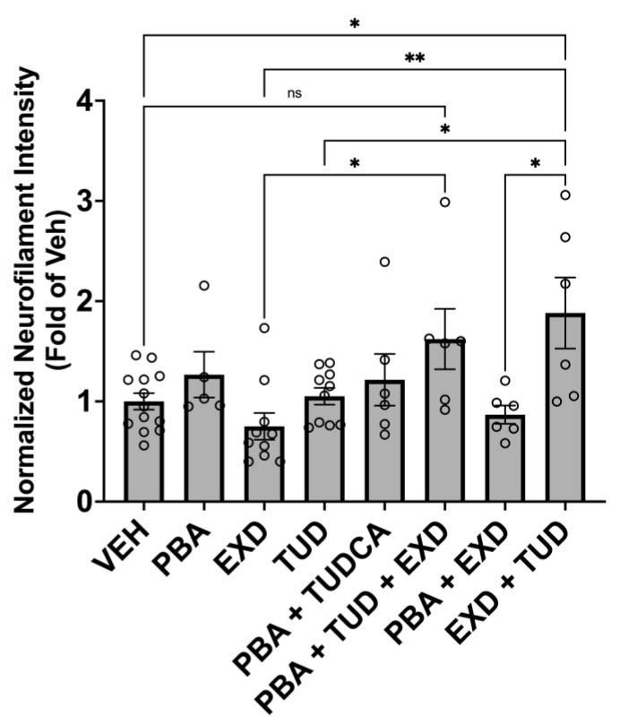
Methods: We evaluated 44 combinations of the nine medications (sodium phenylbutyrate, terazosin, exenatide, ambroxol, deferiprone, coenzyme-Q10, creatine, dasatinib and tauroursodeoxycholic acid) selected for their previously demonstrated evidence of their impact on different targets, showing neuroprotective properties in preclinical models of PD. We utilized wild-type induced pluripotent stem-cell-derived human dopaminergic neurons treated with 1-methyl-4-phenylpyridinium for initial screening. We retested some combinations using an idiopathic PD patient-derived induced pluripotent stem cell line and alpha-synuclein triplication line. We assessed anti-neuroinflammatory effects using human microglia cells. As metrics, we evaluated neurite length, number of branch points per mm2, the number of live neurons, neurofilament heavy chain and pro-inflammatory cytokines.

Results: We have identified four combinations of two to three drugs that showed an additive protective effect in some endpoints. Only the combination of sodium phenylbutyrate, exenatide and tauroursodeoxycholic acid showed improvement in four endpoints studied.

Conclusions: We demonstrated that some of the medications, used in combination, can exert an additive neuroprotective effect in preclinical models of PD that is superior to that of each of the compounds individually. This project can lead to the development of the first treatment for PD that can slow or prevent its progression.

Keywords: Parkinson’s disease; ROS; anti-inflammatory effects; combination therapy; drug repurposing; exenatide; iPSCs; in vitro models; sodium phenylbutyrate; tauroursodeoxycholic acid.


Conflict of interest statement

E.E. and F.S. are from Porsolt Research Laboratory where some of the experiments were performed. D.R. is from Prosoft Clinical and served as a statistician. These companies had no role in the design of the study; or interpretation of data; in the writing of the manuscript, or in the decision to publish the results.

Figures

Figure 1
Figure 1
Multiple processes leading to neurodegeneration in PD include: misfolding of proteins that fail to be cleared from the brain, which in turn stimulates neuroimmune responses; calcium excitotoxicity, affecting mitochondria and resulting in energy depletion, leading to neurodegeneration; iron accumulation that leads to activation of microglia and further neuroinflammation, causing oxidative stress, formation of reactive oxygen species (ROS); mitochondrial dysfunction; and neuroinflammation, all of which ultimately contribute to neurodegeneration. Drugs in parentheses are thought to inhibit the corresponding process. Modified from our previously published diagram [8].
Figure 2
Figure 2
Effect of selected experimental conditions on Area Under Curve (AUC) of neurite length (mm/mm2) (A,C,E,G) and of number of branch points per mm2 (B,D,F,H), 72 h after MPP+ incubation in human dopaminergic iPSCs. The percentage of cytolysis was also determined by evaluating the number of dead cells as compared with the total number of cells at 72 h (I). (a): Compared with Non-Treated conditions (NT); (b): Compared with MPP+ controls (* p < 0.05; ** p < 0.01; *** p < 0.001).
Figure 3
Figure 3
Neurite outgrowth and the number of branches 72 h after incubation with MPP+ and tested drug combinations. Artificial colors are applied on images to show the detection of neurites (pink) and cell bodies (orange) by Incucyte live-cell imaging platform (magnification ×20). (A,D) Non-treated conditions; (B,E) the same cells treated with MPP+ and (C,F) the same cells treated with MPP+ and corresponding drug combinations. PBA: Sodium phenylbutyrate; EXD: Exendin-4; TUDCA: Tauroursodeoxycholic acid.
Figure 4
Figure 4
Effect of experimental conditions on Neurofilament Heavy Chain signal intensity in alpha-synuclein triplication cell line. Line 3x-1 at day 60 postdifferentiation was treated with the indicated compounds for 3 months, then fixed and immunostained for neurofilament protein as an indication of neurite health. Values are the mean +/− SEM, ANOVA with Tukey’s post hoc test (* p < 0.05; ** p < 0.01; ns—not significant). Each plot represents an individual culture well. EXD, Exendin-4; TUD, TUDCA; PBA, sodium phenylbutyrate; VEH, vehicle.
Figure 5
Figure 5
Effects of compound treatment on cellular state of idiopathic PD cell line. (A) Cell count: the cell (nuclei) count is based on the number of Hoechst-stained nuclei per well. Each bar represents the normalized mean cell count per well. (B) Neurite network density: neurites are stained with an antibody against Neurofilament heavy chain, marking all neurites. Each bar represents the mean NFH-positive area per well. For (A,B), the per well values are normalized against the mean of all untreated wells. Error bars represent standard deviation. (* p < 0.05).
Figure 6
Figure 6
Effect of experimental conditions on pro-inflammatory cytokines in human Microglial cells 6 h after LPS incubation (ATP was added 5.5 h after LPS). (A). Effect on IL-6; (B). Effect on TNF-α; (C). Effect on CXCL1/GROα. 1. PBA: Sodium phenylbutyrate at 500 µM; 2. TUDCA at 200 µM; 3. EXD: Exendin at 100 nM; 4. PBA at 500 µM and EXD at 100 nM; 5. PBA at 500 µM and TUDCA at 200 µM; 6. TUDCA at 200 µM and EXD at 100 nM; 7. PBA at 500 µM, TUDCA at 200 µM and EXD at 100 nM; 8. Lower doses of PBA at 100 µM, TUDCA at 40 µM and EXD at 20 nM; 9. Lower doses of PBA at 100 µM and EXD at 20 µM. (a): Compared with Non-Treated conditions (NT); (b): compared with LPS/ATP controls (* p < 0.05; ** p < 0.01; *** p < 0.001).

References

    1. Yang W., Hamilton J.L., Kopil C., Beck J.C., Tanner C.M., Albin R.L., Ray Dorsey E., Dahodwala N., Cintina I., Hogan P., et al. Current and projected future economic burden of Parkinson’s disease in the U.S. npj Park. Dis. 2020;6:15. doi: 10.1038/s41531-020-0117-1. - DOI - PMC - PubMed
    1. Cuervo A.M., Stefanis L., Fredenburg R., Lansbury P.T., Sulzer D. Impaired degradation of mutant alpha-synuclein by chaperone-mediated autophagy. Science. 2004;305:1292–1295. doi: 10.1126/science.1101738. - DOI - PubMed
    1. Mosharov E.V., Larsen K.E., Kanter E., Phillips K.A., Wilson K., Schmitz Y., Krantz D.E., Kobayashi K., Edwards R.H., Sulzer D. Interplay between cytosolic dopamine, calcium, and alpha-synuclein causes selective death of substantia nigra neurons. Neuron. 2009;62:218–229. doi: 10.1016/j.neuron.2009.01.033. - DOI - PMC - PubMed
    1. Ryan S.D., Dolatabadi N., Chan S.F., Zhang X., Akhtar M.W., Parker J., Soldner F., Sunico C.R., Nagar S., Talantova M., et al. Isogenic human iPSC Parkinson’s model shows nitrosative stress-induced dysfunction in MEF2-PGC1alpha transcription. Cell. 2013;155:1351–1364. doi: 10.1016/j.cell.2013.11.009. - DOI - PMC - PubMed
    1. Zucca F.A., Segura-Aguilar J., Ferrari E., Munoz P., Paris I., Sulzer D., Sarna T., Casella L., Zecca L. Interactions of iron, dopamine and neuromelanin pathways in brain aging and Parkinson’s disease. Prog. Neurobiol. 2017;155:96–119. doi: 10.1016/j.pneurobio.2015.09.012. - DOI - PMC - PubMed