. 2025 Apr 11;14(4):456.
doi: 10.3390/antiox14040456.

Effect of 1-DNJ on Oxidative Stress-Induced Apoptosis in Porcine Ovarian GCs Through Modulation of the PERK-ATF4/MFN2 Signaling Pathway

Affiliations

Effect of 1-DNJ on Oxidative Stress-Induced Apoptosis in Porcine Ovarian GCs Through Modulation of the PERK-ATF4/MFN2 Signaling Pathway

Wenwen Xing et al. Antioxidants (Basel). .

Abstract

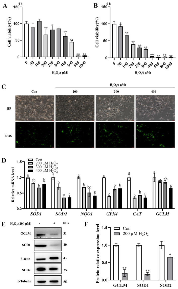
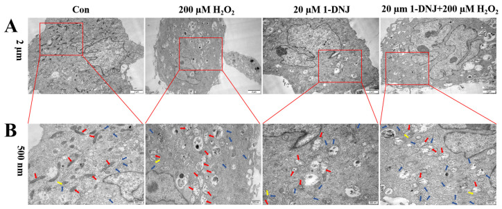
Oxidative stress (OS) is regarded as a major contributor to granulosa cellapoptosis in ovarian disease. 1-Deoxynojirimycin (1-DNJ), a naturally occurring plant alkaloid, exhibits antioxidant, anti-inflammatory, and metabolism-modulating properties. Mitochondria and endoplasmic reticulum (ER), crucial organelles regulating oxidative balance, interact through mitochondria-associated endoplasmic reticulum membranes (MAMs) for signaling and molecular exchange. However, it remains unclear whether 1-DNJ attenuates oxidative damage in ovarian granulosa cells (GCs) via MAMs-mediated ER-mitochondria crosstalk, which needs further exploration. This study aimed to investigate the mechanisms by which 1-DNJ affects oxidative damage and apoptosis induced by OS in porcine follicular GCs by regulating mitochondrial function, MAMs, and ER interactions. Here, we found that GCs suffered from OS, accompanied by the up-regulation of ROS and MDA, alongside reduced activity of antioxidant enzymes (CAT and T-SOD). Further studies revealed that the up-regulation of MAMs proteins (MFN2, MCU, and VDAC1) and pro-apoptosis proteins (BAX and Cleaved-capase3), along with increased mitochondrial ROS and Ca2+ levels, led to the down-regulation of MMP and ATP content. These, in turn, triggered mitochondrial dysfunction, and MAMs destabilization, and subsequent apoptosis. Additionally, the up-regulation of the protein levels of P-PERK/PERK, GRP78, ATF4, and CHOP protein expression activated the PERK-ATF4 signaling pathway, which triggered endoplasmic reticulum stress (ERS). Conversely, 1-DNJ alleviated H2O2-induced mitochondrial and MAMs dysfunction and ERS, which in turn attenuated apoptosis. Further, ATF4 knockdown inhibited MFN2 protein expression, which attenuated H2O2-induced MMP inhibition, Ca2+ overload, ROS production, and mitochondrial damage. In summary, 1-DNJ mitigated OS-induced mitochondrial dysfunction in GCs and regulated ER-mitochondrial communication through MAMs, reducing OS-induced apoptosis. The present study demonstrates that 1-DNJ protects ovarian GCs from OS-induced damage by modulating ER and mitochondrial homeostasis through MAMs, offering new perspectives and a theoretical basis for the treatment of ovarian diseases.

Keywords: 1-DNJ; endoplasmic reticulum stress; mitochondria-associated endoplasmic reticulum membrane; mitochondrial dysfunction; ovarian granulosa cells; oxidative stress.


Conflict of interest statement

No conflicts of interest exist in the submission of this manuscript, and the manuscript is approved by all authors for publication. I would like to declare on behalf of my coauthors that the work described was original research that has not been published previously, and is not under consideration for publication elsewhere, in whole or in part. All of the authors listed have approved the manuscript that is enclosed.

Figures

Figure 1
Figure 1
Modeling of OS in GCs in vitro. (A,B) Cellular activity of GCs after treatment with different concentrations of H2O2 (0, 50, 100, 200, 250, 300, 400, 500, 800, and 1000 µM) for 4 or 6 h, respectively, was determined using the CCK8 kit; (C) a DCFH-DA fluorescent probe was used to detect the ROS accumulation in GCs 6 h after treatment with 0, 200, 300, and 400 µM of H2O2. The scale bar of the graph is 50 µm and the magnification is 400×. (D) qRT-PCR was used to detect mRNA expression of OS-related genes in GCs induced by 0, 200, 300, and 400 µM of H2O2 for 6 h; (E,F) Western blot was used to detect the protein expression levels of GCLM, SOD1, and SOD2 in GCs induced by 0 and 200 µM H2O2 for 6 h. a–c There are no significant differences between groups with the same superscript letter, while there are significant differences between groups with different lowercase superscript letters (p < 0.05, ANOVA). * p < 0.05 and ** p < 0.01 compared with the Con group (Student’s t-test).
Figure 2
Figure 2
Effect of 1-DNJ on cell viability of GCs. Cellular viability of GCs treated with different concentrations of 1-DNJ (0, 5, 10, 20, 30, 40, 50, 70, and 80 µM) for 24 h was determined using the CCK8 kit. * p < 0.05 and ** p < 0.01 compared with 0 µM of 1-DNJ (Student’s t-test).
Figure 3
Figure 3
1-DNJ antioxidant analysis in GCs. (A) qRT-PCR was used to detect the mRNA expression of OS-related genes in GCs treated with 0, 10, 20, and 30 µM of 1-DNJ for 24 h; (B,C) Western blot detection of SOD1, SOD2, and GCLM protein expression levels of GCs at 0 and 20 µM of 1-DNJ treatment for 24 h. a–c There are no significant differences between groups with the same superscript letter, while there are significant differences between groups with different lowercase superscript letters (p < 0.05, ANOVA). Compared with the Con group, * p < 0.05 and ** p < 0.01 (Student’s t-test).
Figure 4
Figure 4
1-DNJ inhibits ROS release and enhances antioxidant defense of GCs. GCs were first treated with 1-DNJ (20 µM) for 24 h and then with H2O2 (200 µM) for 6 h. (A) ROS accumulation in GCs was detected by using a DCFH-DA fluorescent probe. The scale bar was 200 µm and the magnification was 100×. (B) CAT and T-SOD activities and MDA content of GCs were determined using kits, respectively. (C) qRT-PCR was used to detect mRNA expression of OS-related genes in GCs. (D,E) Western blot was performed to detect the protein expression levels of SOD1, SOD2, and GCLM in GCs. a–d There are no significant differences between groups with the same superscript letter, while there are significant differences between groups with different lowercase superscript letters (p < 0.05, ANOVA). Compared with the Con group, * p < 0.05 (Student’s t-test); compared with the H2O2 group, # p < 0.05 and ## p < 0.01 (Student’s t-test).
Figure 5
Figure 5
Effect of 1-DNJ on cell viability reduced by H2O2. The GCs were first pretreated with 20 µM 1-DNJ for 24 h, and then induced with 200 and 300 µM, respectively, of H2O2 for 6 h. The cell activity was determined using the CCK8 kit. Compared with the Con group, ** p < 0.01 (Student’s t-test); compared with the H2O2 group,## p < 0.01 (Student’s t-test).
Figure 6
Figure 6
Structures of mitochondria and ER ultrastructure within GCs collected under transmission electron microscopy. GCs were first treated with 1-DNJ (20 µM) for 24 h and then with H2O2 (200 µM) for 6 h. Red arrows indicate mitochondria; blue arrows indicate ER, yellow arrows indicate MAMs. Scale bars: (A) is 2 µm and (B) is 500 nm.
Figure 7
Figure 7
Effect of 1-DNJ on H2O2-induced mitochondrial dysfunction in GCs. GCs were first treated with 1-DNJ (20 µM) for 24 h and then with H2O2 (200 µM) for 6 h. (A) ATP content in GCs was detected using an ATP assay kit. (B,C) The content of NAD+ in GCs and the NAD+/NADH ratio was detected using an NAD+/NADH assay kit. (D) Changes in MMP in GCs were assessed with the fluorescent probe JC-1. (E) Detection of mtROS levels within GCs was assessed using the MitoSOX Red fluorescent probe. (D,E) were scaled to 100 µm and magnified to 200×. a–d There are no significant differences between groups with the same superscript letter, while there are significant differences between groups with different lowercase superscript letters (p < 0.05, ANOVA).
Figure 8
Figure 8
Effect of 1-DNJ on OS-induced apoptosis of GCs. GCs were first treated with 1-DNJ (20 µM) for 24 h and then with H2O2 (200 µM) for 6 h. (A) qRT-PCR was used to detect the mRNA expression of apoptosis-related genes. (B,C) Western blot was used to detect the expression levels of apoptotic proteins: BAX, Bcl2, and Cleaved-caspase3. a–c There are no significant differences between groups with the same superscript letter, while there are significant differences between groups with different lowercase superscript letters (p < 0.05, ANOVA). Compared with the Con group, * p < 0.05 (Student’s t-test).
Figure 9
Figure 9
Effect of 1-DNJ on the function of MAMs in GCs by OS. GCs were first treated with 1-DNJ (20 µM) for 24 h and then with H2O2 (200 µM) for 6 h. (A) Ca2+ content in GCs was measured using the fluorescent probe Rhod-2/AM, the scale bar was 100 µm and the magnification was 200 ×. (B,C) Western blot was used to detect the expression levels of MAMs-representative proteins MCU, MFN2, and VDAC1. a–c There are no significant differences between groups with the same superscript letter, while there are significant differences between groups with different lowercase superscript letters (p < 0.05, ANOVA).
Figure 10
Figure 10
Effect of 1-DNJ on OS-induced endoplasmic reticulum stress response in GCs. GCs were first treated with 1-DNJ (20 µM) for 24 h and then with H2O2 (200 µM) for 6 h. (A) Changes in ATF4 expression in GCs were detected by immunofluorescence; (B,C) Western blot detection of p-PERK/PERK, GRP78, ATF4, and CHOP protein expression; and (D) qRT-PCR detection of GRP78, ATF4, and CHOP mRNA expression. (E) Molecular docking simulation for the ligand−protein binding of 1-DNJ with ATF4. (A) Scale bar is 200 µm and magnification is 100×. a–d There are no significant differences between groups with the same superscript letter, while there are significant differences between groups with different lowercase superscript letters (p < 0.05, ANOVA). Compared with the Con group, * p < 0.05 (Student’s t-test).
Figure 11
Figure 11
Crosstalk between ERS, OS, and mitochondrial dysfunction. GCs were first treated with 1-DNJ (20 µM) for 24 h, followed by the transfection of ATF4 knockdown for 24 h, and then exposed to H2O2 (200 µM) for 6 h. (A) MMP changes within GCs were detected using JC-1, the fluorescent probe. (B) Ca2+ content in GCs was measured using the Rhod-2/AM fluorescent probe. (C) ROS accumulation in GCs was detected using the DCFH-DA fluorescent probe. (D) WB analysis was used to detect the expression levels of MAMs-representative proteins. (AC) The scale bar is 100 µm and the magnification is 200×. a–d There are no significant differences between groups with the same superscript letter, while there are significant differences between groups with different lowercase superscript letters (p < 0.05, ANOVA). Compared with the NC + H2O2 group, ** p < 0.01 (Student’s t-test).
Figure 12
Figure 12
Mechanism of 1-DNJ alleviating OS in porcine GCs. (A) When OS occurs in porcine GCs, the ERS pathway is activated, leading to ER damage and the triggering of ERS. Meanwhile, mitochondrial function is impaired and the level of mitochondrial OS is increased. During this process, OS affects the communication between the mitochondria and ER through MAMs, resulting in an increase in the content of Ca2+ in the mitochondria and changes in the activities of apoptosis-related proteins Bax, Bcl2, and Caspase3. Ultimately, this leads to an increase in apoptosis of porcine GCs. (B) When OS occurs in GCs, the addition of 1-DNJ can inhibit the activity of the PERK-ATF4 signaling pathway, relieve ERS, restore ER and mitochondrial functions, prevent ER calcium inflow from the ER into the mitochondria by regulating the expression of MAMs-related proteins and MCU, reduce mitochondrial OS levels, and restore MMP and ATP synthesis. Moreover, MFN2 is a potential target gene downstream of ATF4. Ultimately, 1-DNJ promotes the expression of the anti-apoptosis-related protein Bcl2, inhibits the expression of pro-apoptosis proteins BAX and Caspase3, and finally, alleviates the apoptosis of porcine GCs caused by OS. This figure was drawn by Figdraw.

References

    1. Duan H., Yang S., Yang S., Zeng J., Yan Z., Zhang L., Ma X., Dong W., Zhang Y., Zhao X., et al. The mechanism of curcumin to protect mouse ovaries from oxidative damage by regulating AMPK/mTOR mediated autophagy. Phytomedicine. 2024;128:155468. - PubMed
    1. Wang Y., Pattarawat P., Zhang J., Kim E., Zhang D., Fang M., Jannaman E.A., Yuan Y., Chatterjee S., Kim J.J., et al. Effects of Cyanobacterial Harmful Algal Bloom Toxin Microcystin-LR on Gonadotropin-Dependent Ovarian Follicle Maturation and Ovulation in Mice. Environ. Health Perspect. 2023;131:67010. - PMC - PubMed
    1. Lu J., Wang Z., Cao J., Chen Y., Dong Y. A novel and compact review on the role of oxidative stress in female reproduction. Reprod. Biol. Endocrinol. 2018;16:80. - PMC - PubMed
    1. Wang X., Yang J., Li H., Mu H., Zeng L., Cai S., Su P., Li H., Zhang L., Xiang W. miR-484 mediates oxidative stress-induced ovarian dysfunction and promotes granulosa cell apoptosis via SESN2 downregulation. Redox Biol. 2023;62:102684. - PMC - PubMed
    1. Huang L., Yuan H., Shi S., Song X., Zhang L., Zhou X., Gao L., Pang W., Yang G., Chu G. CLOCK inhibits the proliferation of porcine ovarian granulosa cells by targeting ASB9. J. Anim. Sci. Biotechnol. 2023;14:82. - PMC - PubMed

LinkOut - more resources