Review
. 2025 Apr 15;14(4):471.
doi: 10.3390/antiox14040471.

Major Oxidative and Antioxidant Mechanisms During Heat Stress-Induced Oxidative Stress in Chickens

Affiliations
Review

Major Oxidative and Antioxidant Mechanisms During Heat Stress-Induced Oxidative Stress in Chickens

Bikash Aryal et al. Antioxidants (Basel). .

Abstract

Heat stress (HS) is one of the most important stressors in chickens, and its adverse effects are primarily caused by disturbing the redox homeostasis. An increase in electron leakage from the mitochondrial electron transport chain is the major source of free radical production under HS, which triggers other enzymatic systems to generate more radicals. As a defense mechanism, cells have enzymatic and non-enzymatic antioxidant systems that work cooperatively against free radicals. The generation of free radicals, particularly the reactive oxygen species (ROS) and reactive nitrogen species (RNS), under HS condition outweighs the cellular antioxidant capacity, resulting in oxidative damage to macromolecules, including lipids, carbohydrates, proteins, and DNA. Understanding these detrimental oxidative processes and protective defense mechanisms is important in developing mitigation strategies against HS. This review summarizes the current understanding of major oxidative and antioxidant systems and their molecular mechanisms in generating or neutralizing the ROS/RNS. Importantly, this review explores the potential mechanisms that lead to the development of oxidative stress in heat-stressed chickens, highlighting their unique behavioral and physiological responses against thermal stress. Further, we summarize the major findings associated with these oxidative and antioxidant mechanisms in chickens.

Keywords: antioxidant mechanisms; chickens; heat stress; oxidative stress; reactive oxygen species.


Conflict of interest statement

The authors declare no conflicts of interest.

Figures

Figure 1
Figure 1
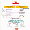
Heat stress (HS)-induced oxidative stress and the indicators of oxidative stress. Primary contributors to cellular reactive oxygen species (ROS) and reactive nitrogen species (RNS) generation include mitochondrial electron transport chain (ETC), NADPH oxidase (NOX), xanthine oxidase (XO), and uncoupled endothelial nitric oxide synthase (eNOS). Heat stress primarily triggers mitochondrial ROS production, which then activates other enzymatic systems, showing significant interplay between them. Increased levels of mitochondrial ROS disrupt the endoplasmic reticulum (ER) homeostasis, leading to the buildup of misfolded proteins, which in turn prompt mitochondria to produce more ROS. The resulting feed-forward mechanisms amplify ROS production and lead to the oxidative damage of DNA, lipids, proteins, and carbohydrates.
Figure 2
Figure 2
Heat stress (HS)-induced reactive oxygen species (ROS) production in mitochondria. Electrons transport through complex-I, -III, and -IV drives proton transfer to intermembrane space, creating an electrochemical proton gradient that ultimately moves protons back through ATP synthase to generate ATP. Uncoupling protein (UCP), through proton leakage, uncouples oxidative phosphorylation and regulates mitochondrial ROS production. Heat stress increases energy metabolisms, affecting the enzymatic activity of respiratory chain complexes. This alters the UCP expression as well as increases electron leakage from complex-I and complex-III, resulting in superoxide (O2•−) production, which can have several fates. Mitochondrial manganese superoxide dismutase (Mn-SOD) catalyzes the conversion of O2•− to hydrogen peroxide (H2O2), which can be reduced to water by several cellular enzymes, including the glutathione peroxidase (GPx) and catalase (CAT). H2O2 can also result in the generation of hydroxyl radical (HO) with the availability of transition metals through Fenton chemistry or by reacting with the O2•− via Haber–Weiss reaction. Rapid inactivation of nitric oxide (NO) in the presence of O2•− resulted in the yield of peroxynitrite (ONOO), a strong oxidant that subsequently undergoes protonation or decomposes into other reactive species, such as carbonate radical (CO3•−) and nitrogen dioxide radical (NO2).
Figure 3
Figure 3
Endoplasmic reticulum (ER) stress and mitochondrial reactive oxygen species (ROS) production exhibit a positive feed-forward loop. In an attempt to mitigate the accumulation of unfolded proteins and ER stress, three ER transmembrane proteins, inositol requiring 1 (IRE1), PKR-like ER kinase (PERK), and activating transcription factor 6 (ATF6), in the ER lumen are triggered and activate a set of signaling cascades called the unfolded protein response (UPR). The ER represents the primary storage site of calcium (Ca2+), and under ER stress, Ca2+ from ER cisternae flows through the calcium release channels, inositol 1,4,5-trisphosphate receptors (IP3R) and ryanodine receptors (RyR). The Ca2+ thus released from the ER is sequestered by mitochondria through mitochondria-associated ER membranes (MAMs). Calcium overload in mitochondria triggers ROS production by increasing the respiratory chain activity and opening the mitochondrial permeability transition pore (mPTP), causing the release of cytochrome C and other pro-apoptotic factors. This, in turn, exacerbates ER stress, further releasing Ca2+ and optimizing mitochondrial ROS production.
Figure 4
Figure 4
Potential downstream mechanisms associated with the initial responses, hyperventilation, and wings flutter, against heat stress: (a) Panting, as a way of evaporative cooling, leads to hyperventilation. Excessive loss of carbon dioxide (CO2) develops in respiratory alkalosis, which could eventually lead to metabolic acidosis through decreasing hydrogen ion (H+) secretion while increasing bicarbonate ion (HCO3) secretion and chloride ion (Cl) reabsorption from the kidney. (b) On the other hand, as a result of increased peripheral circulation, the blood supply to the internal organs is reduced, creating a hypoxic environment. Cells metabolize glucose anaerobically under hypoxia, resulting in the production and accumulation of lactic acid. The resultant acidosis condition stimulates mitochondria to uptake more calcium, which increases metabolic activity and subsequent reactive oxygen species (ROS) production. Acidosis can also alter oxidative phosphorylation by reducing the signal for ATP synthesis (i.e., ADP), and by modulating the mitochondrial fatty acids oxidation that decreases complex-I activity by protein acetylation. All of these mechanisms result in increased ROS generation.
Figure 5
Figure 5
Potential downstream mechanisms associated with the initial response, stress hormone, against heat stress. The conventional and well-established mode of action of glucocorticoid (GC) is through binding to its receptor, glucocorticoid receptors (GRs). Upon nuclear localization and binding to its genomic response element (GRE), the GC-GR complex exhibits various anti-inflammatory and metabolic effects, including the deficiency of GTP cyclohydrolase I (GCH1) and downregulation of cationic amino acid transporter 1 (CAT-1). This, in turn, decreases the bioavailability of tetrahydrobiopterin (BH4) and L-arginine respectively, leading to nitric oxide synthase (NOS) uncoupling. Moreover, GR associated with heat shock protein (HSP) in mitochondria forms a complex with the anti-apoptotic protein B-cell lymphoma 2 (BCL2). Chronic exposure to the GC downregulates the binding of GR with the Bcl-2, which leads to the formation of Bax pores and subsequent leakage of calcium ions and cytochrome c. The GC also exerts its rapid non-genomic effects, such as NADPH oxidase (NOX) activation, via modulation of various kinases. Therefore, the combined effects of mitochondrial dysfunction, NOS uncoupling, and NOX activation ultimately lead to a rise in reactive oxygen species (ROS) production.
Figure 6
Figure 6
Potential downstream mechanisms associated with the initial response, decreased feed intake, against heat stress. Energy deficit condition activates lipolysis and subsequent release of free fatty acids (FFAs), with polyunsaturated fatty acids (PUFAs) activating NADPH oxidase (NOX) and saturated fatty acids (SFAs) triggering endoplasmic reticulum (ER) stress. The subsequent beta-oxidation of FFAs, especially the SFAs, increases the flavin adenine dinucleotide and reduced nicotinamide adenine dinucleotide ratio (FADH2:NADH), which reduces the number of electron acceptors for complex-I, ultimately increasing electron leakage and reactive oxygen species (ROS) production. Excessive acetyl-CoA generated from beta-oxidation increases the acetylation of mitochondrial proteins via both enzymatic and non-enzymatic mechanisms, which further reduces complex-I activity. The decreased NAD+:NADH ratio resulting from complex-I defect suppresses the deacetylase sirtuin 3 (SIRT3) enzyme, aggravating protein acetylation and ROS production. The cytochrome-C that transfers electrons from complex-III to -IV is released from the mitochondria as a result of ROS accumulation, finally inducing apoptosis.
Figure 7
Figure 7
Schematic representation of major enzymatic and non-enzymatic antioxidant defense systems. Superoxide dismutase (SOD) is the forefront antioxidant that neutralizes superoxide (O2•−) to hydrogen peroxide (H2O2), which is then detoxified to water by catalase (CAT) and the thiol–disulfide system, involving glutathione peroxidase (GPx) and peroxiredoxin (PrDx). The antioxidant network refers to the synergistic relationship between different antioxidants such as vitamin E, vitamin C, and glutathione (GSH), where one replenishes the original properties of another. Vitamin E scavenges lipid peroxyl radicals (LOO), peroxyl radicals (ROO), and hydroxyl radicals (HO) via a chain breaking mechanism and protects the cellular membranes. Cellular GSH, synthesized endogenously from methionine and cysteine, and cellular thioredoxin (Trx) are the important components of the thiol–disulfide system and the antioxidant network. Nicotinamide adenine dinucleotide phosphate (NADPH) serves as a reducing agent by donating electrons to oxidized Trx via thioredoxin reductase (TrxR), or to oxidized glutathione (GSSG) via glutathione reductase (GR).
Figure 8
Figure 8
Thiol–disulfide-mediated redox cycle. By donating electrons to reactive oxygen species (ROS), thiols (R-SH) function as reducing agents to neutralize ROS into less toxic byproducts, while they become oxidized to form disulfides (R-S-S-R). Conversion of thiols to disulfide is mediated by a variety of protein disulfide isomerase (PDI) oxidases, including glutathione peroxidase (GPx) and peroxiredoxin (PrDx). Specific reductase then restores disulfides to reduced thiols by utilizing the cellular reducing agents, including NADPH. This interconversion between thiols and disulfide represents the redox cycle.
Figure 9
Figure 9
The thioredoxin (Trx) system and glutathione (GSH) systems in regulating the thiol–disulfide-mediated redox state of the cell. Both systems utilize NADPH as a final electron donor; however, they operate via different components in reducing the proteins and neutralizing hydrogen peroxide (H2O2) to water. The Trx system includes thioredoxin reductase (TrxR), thioredoxin (Trx), and peroxiredoxin (PrDx) while the GSH system comprises glutathione reductase (GR), glutathione (GSH and GSSG), and glutathione peroxidase (GPx) or glutaredoxin (Grx).
Figure 10
Figure 10
Methionine metabolism, transsulfuration pathway, and the cysteine catabolic pathways. Methionine is an essential amino acid that is converted to homocysteine through the transmethylation pathway. Homocysteine can then be reconverted back into methionine via remethylation pathways, involving a methyl donation from the folate cycle or the betaine. Methionine can also be regenerated via the salvage pathway involving the recycling of methylthioadenosine (MTA), a by-product of polyamine biosynthesis. Alternatively, homocysteine can be converted into cysteine via the transsulfuration pathway. Cysteine has multiple fates and is catabolized to produce various nonprotein compounds, such as glutathione (GSH), taurine, sulfate, and coenzyme A depending on the physiological and nutritional situations.
Figure 11
Figure 11
Schematic representation of the ubiquitin–proteasome system (UPS) and the molecular chaperones in regulating proteostasis. Misfolded protein resulting from oxidative injury activates the UPS, leading to the ubiquitination of misfolded proteins and 26S proteasomal degradation. The primary role of molecular chaperones, including the heat shock protein 70 (HSP70) and heat shock protein 90 (HSP90), is to refold the misfolded proteins; however, they have also been demonstrated to assist in the degradation of misfolded proteins through the UPS. In addition, HSPs play roles in protein assembly/disassembly, their translocation across membranes, and activity regulation of secretory proteins.
Figure 12
Figure 12
Schematic representation of the nuclear factor erythroid 2-related factor 2 (Nrf2) pathway and heat shock factor 1 (HSF1) pathway in regulating oxidative stress and proteotoxic stress, respectively: (a) Under normal conditions, kelch-like ECH-associated protein 1 (Keap1) binds with Nrf2, which will be subsequently ubiquitinated by Cullin 3 (Cul3) ligase for proteasomal degradation. During oxidative stress, reactive oxygen species (ROS) modifies cysteine residues in Keap1, leading to Nrf2 stabilization, nuclear translocation, and target gene binding to trigger the expression of multiple antioxidant genes. (b) HSF, particularly HSF1, is activated by both proteotoxic stimuli such as heat shock and oxidative stress. Upon activation, it trimerizes and interacts with the heat shock element (HSE) of its target genes in the nucleus, thereby regulating the expression of heat shock proteins (HSPs). As a molecular chaperone, HSPs assist in proper protein folding and mitigate proteotoxic stress.
Figure 13
Figure 13
Schematic representation of the nuclear factor kappa B (NF-κB) pathway and mitogen-activated protein kinase (MAPK) pathway in regulating oxidative stress: (a) Reactive oxygen species (ROS)-induced oxidative stress has a dual role in NF-κB activity; the early phase activates the NF-κB pathway while prolonged exposure can inhibit the degradation of the inhibitor of nuclear factor kappa B (IκB), thus preventing NF-κB activation. Additionally, the NF-κB pathway can exert both antioxidant as well as pro-oxidant functions during the time of oxidative stress by regulating the expression of various antioxidant genes and pro-oxidant genes, respectively. (b) Activation of another transcription factor, activator protein-1 (AP-1), is linked with cellular proliferation, inflammation, and death. AP-1 is the primary target of MAPK, which has three subfamilies, namely, extracellular signal-regulated kinase (ERK), c-Jun N-terminal kinase (JNK), and p38. One mechanism by which oxidative stress triggers MAPK and AP-1 is the inactivation and degradation of mitogen-activated protein kinase phosphatase (MKP). Under normal conditions, MKP negatively regulates MAPK by dephosphorylation, thereby rendering its activity. However, ROS can promote the degradation of MKP, resulting in the activation of MAPK signaling.

Similar articles

Cited by

References

    1. Sisein E.A. Biochemistry of free radicals and antioxidants. Sch. Acad. J. Biosci. 2014;2:110–118.
    1. Martemucci G., Costagliola C., Mariano M., D’andrea L., Napolitano P., D’Alessandro A.G. Free radical properties, source and targets, antioxidant consumption and health. Oxygen. 2022;2:48–78. doi: 10.3390/oxygen2020006. - DOI
    1. Pham-Huy L.A., He H., Pham-Huy C. Free radicals, antioxidants in disease and health. Int. J. Biomed. Sci. 2008;4:89. doi: 10.59566/IJBS.2008.4089. - DOI - PMC - PubMed
    1. Halliwell B. eLS. Wiley; Hoboken, NJ, USA: 2001. Free radicals and other reactive species in disease.
    1. Sies H., Jones D.P. Reactive oxygen species (ROS) as pleiotropic physiological signalling agents. Nat. Rev. Mol. Cell Biol. 2020;21:363–383. doi: 10.1038/s41580-020-0230-3. - DOI - PubMed

LinkOut - more resources