. 2025 May;36(5):1095-1103.
doi: 10.1007/s00192-025-06083-4. Epub 2025 Apr 29.

Feasibility of the CT-Image-Guided Colpopexy: A Novel, Needle-Based Sacrospinous Ligament Fixation for Treatment of Vaginal Apical Prolapse

Affiliations

Feasibility of the CT-Image-Guided Colpopexy: A Novel, Needle-Based Sacrospinous Ligament Fixation for Treatment of Vaginal Apical Prolapse

Boram Choi et al. Int Urogynecol J. 2025 May.

Abstract

Introduction and hypothesis: Although several procedures have been developed to increase the precision and reduce the invasiveness of sacrospinous ligament fixation surgery, all surgical approaches require dissection of the vagina to access the paravaginal space and visual or tactile identification of the ligament before deploying a suture or anchor device at the ligament target site. The aim of this study was to develop and demonstrate the feasibility of a minimally invasive, needle-based treatment of vaginal apical prolapse by CT-image-guided anchoring of the vaginal vault to the sacrospinous ligament in cadavers.

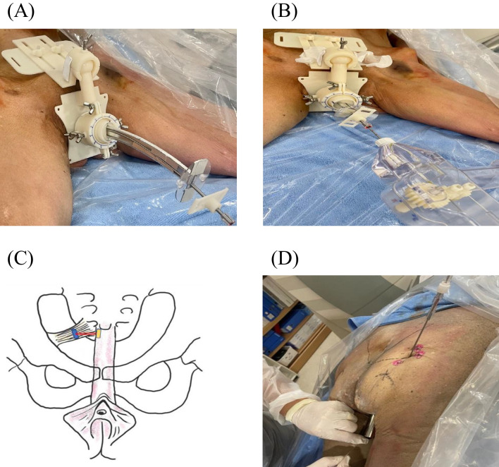
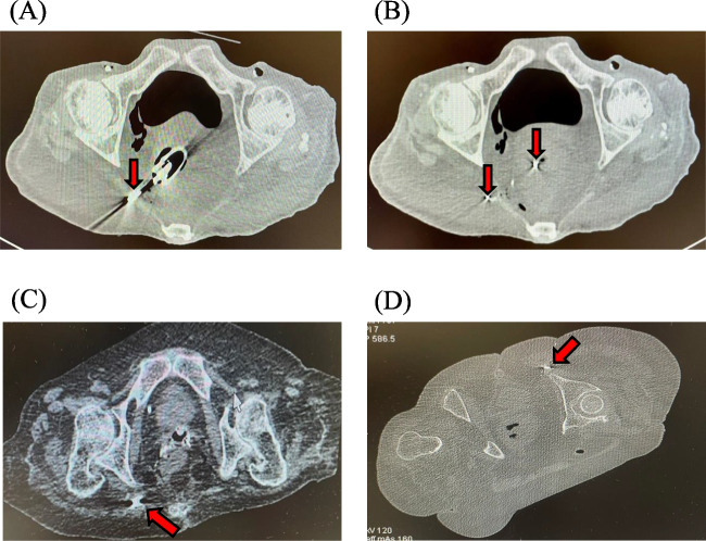
Methods: A CT-image-guided, needle-based colpopexy procedure was performed on six female cadavers. After the placement of the vaginal probe, each cadaver was CT scanned to identify the target sacrospinous ligament. An anchoring device was inserted through the vaginal probe toward the target ligament. Small anchors, introduced through needle lumens via either transvaginal or transgluteal access, were implanted under the guidance of CT images to anchor the vaginal vault to the sacrospinous ligament. The cadavers were dissected and compared with CT images to evaluate the anatomical locations of the anchors with respect to the anatomical landmarks in the pelvis. Pull-out forces of the implants were also measured.

Results: Thirty-two anchors were inserted via transvaginal access, whereas ten anchors were inserted via transgluteal access. CT images revealed that the inserted anchors were implanted in the sacrospinous ligaments (n = 40: 30 transvaginal and 10 transgluteal) or sacrotuberous ligaments (n = 2: transvaginal), as confirmed by dissection of the gluteal region of the cadavers. The mean pullout force was measured as 47.5 N ± 5.0.

Conclusions: We report a feasibility study for the application of novel needle-based colpopexy under CT-image guidance. Further clinical studies are required to implement this method in clinical settings and to demonstrate its safety and effectiveness compared with conventional surgical procedures.

Keywords: CT; Cadaver; Image-guided intervention; Medical device; Minimally invasive procedure; Pelvic organ prolapse.


Conflict of interest statement

Declarations. Ethical/Institutional Review Board Approval: Catholic University of Korea Seoul St. Mary’s Hospital IRB Review Result Notice [Form Number: MC22EIGC0074]. Conflicts of Interest: B.C., K.H.L., and H.C. have no conflicts of interest to declare. K.T.B. and C.K. submitted a provisional patent application.

Figures

Fig. 1
Fig. 1
Schematic perspective and transverse cross-sectional drawings of female pelvic anatomy illustrating sacrospinous ligament fixation (SSLF) procedures. A, B The transvaginal approach to SSLF involves the placement of a vaginal probe and a needle projected (the direction denoted by the arrow) from the opening port of the vaginal probe to the target SSL in a patient in the supine position. C, D The transgluteal approach of SSLF involves the placement of a vaginal probe and a needle projected (the direction denoted by the arrow) from a subcutaneous site to the vaginal vault via the target SSL in a patient in the prone position
Fig. 2
Fig. 2
Vaginal probe and anchoring device. A Images of the vaginal probe used in the transvaginal approach. B Image of an anchoring device used for both the transvaginal and the transgluteal approaches. C 3D-rendered perspective drawing of an anchoring device inserted through a vaginal probe for the transvaginal approach
Fig. 3
Fig. 3
Drawings of a transgluteal vaginal probe with a needle inserted, a needle loaded with a retainer, and a retainer with two anchors apposed to the walls of the ligament and vagina. A 3D-rendered perspective drawing of a vaginal probe for the transgluteal approach with a needle inserted into the distal port of the vaginal probe. B Magnified drawing of the vaginal probe for the transgluteal approach with an inserted needle, through which a retainer anchor partially exposed the retention wire into the distal port of the vaginal probe. C Cross-sectional view of a needle loaded with a retainer consisting of two anchors and a retention wire. D Illustration of the two anchors of a retainer apposed to the walls of the ligament and vagina, which are fastened by the retention wire
Fig. 4
Fig. 4
Cadavers on a CT scanner table undergoing the sacrospinous ligament fixation (SSLF) procedure and a schematic perspective drawing showing the SSLF outcome. A A transvaginal vaginal probe with a stabilizer was placed over the pelvis prior to advancement of the vaginal probe into the vaginal lumen. B An anchoring device was inserted through the transvaginal probe. C A schematic perspective drawing of the female pelvic anatomy showing the vaginal vault fastened to the right SSL using anchors and retention wire. D A transgluteal vaginal probe is placed in the vagina, toward which a needle is inserted from the right buttock
Fig. 5
Fig. 5
Pelvic CT images of three cadavers undergoing sacrospinous ligament fixation (SSLF) procedures. A Cadaver #6, placed in the supine position for a transvaginal approach to SSLF, shows a vaginal probe in the vagina and a needle (arrow) directed to the right SSL. Black and white linear streaky artifacts from metallic objects were observed on the CT image. B Cadaver #6 shows two anchors (arrows) of a retainer: the proximal anchor in the vagina connected to the distal anchor at the right SSL (i.e., achieving colpopexy). C Cadaver #3, placed in the supine position for a transvaginal approach to SSLF, shows an anchor (arrow) of a retainer placed at the right SSL. D Cadaver #3, placed in the slightly oblique prone position for a transgluteal approach to SSLF, shows an anchor (arrow) of a retainer placed at the left SSL
Fig. 6
Fig. 6
Anchors are anatomically located in association with neighboring pelvic structures during dissection. A Anchor (arrow) at the sacrospinous ligament and (B) anchor (arrow) at the sacrotuberous ligament. Gmax gluteus maximus muscle, P piriformis muscle, STL sacrotuberous ligament

Similar articles

References

    1. Olsen AL, Smith VJ, Bergstrom JO, Colling JC, Clark AL. Epidemiology of surgically managed pelvic organ prolapse and urinary incontinence. Obstet Gynecol. 1997;89:501–6. - PubMed
    1. Wu JM, Vaughan CP, Goode PS, Redden DT, Burgio KL, Richter HE, Markland AD. Prevalence and trends of symptomatic pelvic floor disorders in U.S. women. Obstet Gynecol. 2014;123:141–8. - PMC - PubMed
    1. Wu JM, Hundley AF, Fulton RG, Myers ER. Forecasting the prevalence of pelvic floor disorders in U.S. women: 2010 to 2050. Obstet Gynecol. 2009;114:1278–83. - PubMed
    1. Yazdany T, Wong K, Bhatia NN. Sacrospinous ligament fixation for pelvic organ prolapse in the era of vaginal mesh kits. Curr Opin Obstet Gynecol. 2011;23:391–5. - PubMed
    1. Schulten SFM, Detollenaere RJ, Stekelenburg J, IntHout J, Kluivers KB, van Eijndhoven HWF. Sacrospinous hysteropexy versus vaginal hysterectomy with uterosacral ligament suspension in women with uterine prolapse stage 2 or higher: observational follow-up of a multicentre randomised trial. BMJ. 2019;366:l5149. - PMC - PubMed

LinkOut - more resources