. 2025 Jun 1;328(6):C1880-C1892.
doi: 10.1152/ajpcell.00822.2024. Epub 2025 Apr 29.

Orai channel pharmacological manipulation reduces metabolic flexibility in cardiac fibroblasts

Affiliations

Orai channel pharmacological manipulation reduces metabolic flexibility in cardiac fibroblasts

Patricia da Silva Pantoja Newman et al. Am J Physiol Cell Physiol. .

Abstract

Cardiac fibroblasts (CFs) play a crucial role in regulating normal heart function and are also involved in the pathological remodeling of the heart that occurs due to hypertension, myocardial infarction, and heart failure. Metabolic changes in fibroblasts are key drivers in the progression of these diseases. Calcium (Ca2+) signaling and Ca2+ ion channels control many functions of fibroblasts. Orai Ca2+ channels are abundantly expressed in fibroblasts; however, their exact role is not yet fully understood. This study examined the role of Orai Ca2+ channels in maintaining Ca2+ homeostasis within organelles and in energy production in CFs. We found that chronic inhibition of Orai activity altered the expression levels of major metabolic enzymes, affecting the overall cell metabolism. Orai channels are required to refill the endoplasmic reticulum (ER) store. Acute Orai channel activity inhibition reduced Ca2+ content in the ER and mitochondria and was associated with the impaired ability to use glucose as a primary energy source. These results have significant implications for understanding the role of Orai-dependent Ca2+ entry in maintaining organellar Ca2+ homeostasis and cellular metabolic flexibility, sparking further research in this area.NEW & NOTEWORTHY We show that Orai actively contributes to organellar Ca2+ concentration and energy homeostasis of the cardiac fibroblast. These findings can have a significant impact during fibrogenesis.

Keywords: Orai; calcium; fibroblast; fibrosis; metabolism.


Conflict of interest statement

DISCLAIMERS

No conflicts of interest, financial or otherwise.

Figures

Figure 1.
Figure 1.
Orai inhibition effects on SOCE and CFs proliferation. A) Representative traces from fura-2 loaded cells pre-treated with Tg (10 μM) to activate Orai channels (this portion is not shown), and then Ca2+ is reintroduced in the cell bath to induce SOCE. Fibroblasts preincubated with Orai blockers (2 μM, 5min) exhibited a significantly reduced SOCE compared to vehicle-only treated cells (Ctrl, DMSO 0.2%). B) Orai1 KO fibroblasts pre-treated with Tg (10 μM) exhibit reduction of SOCE as compared to wild-type (WT) fibroblasts. C) Maximal SOCE taken after the addition of CaCl2 (6 mM) to the external medium. D) Maximal SOCE in WT and Orai1 KO fibroblasts taken after the addition of CaCl2 (6 mM) to the external medium. Data generated for each experiment were considered non-paired. Values from each cell from five independent experiments are displayed. For each experiment, the median values are calculated and reported. E) Incucyte® proliferation assay analysis of WT CFS cultured in the presence or absence of different concentrations of CM4620 (0, 2, 5, 10 μM) or vehicle only (DMSO, 0.2%). F) Incucyte® proliferation assay analysis of Orai1 KO CFS cultured in the presence or absence of Orai inhibitor CM4620 (0, 2, 5, 10 μM) or vehicle only (DMSO, 0.2%). G) Images extracted from the Incucyte® proliferation acquisition system after 40 hours post-seeding and treatment with CM4620 (0, 10 μM) or vehicle only (DMSO, 0.2%), arrowheads indicate drug/vehicle addition to the culture. H) Images extracted from the Incucyte® proliferation acquisition system after 40 hours post-seeding and treatment with CM4620 (0, 10 μM) or vehicle only (DMSO, 0.2%). Cells were seeded at 2000 cells per well in a 96-well plate and cultured in the presence or absence of Orai inhibitor CM4620. Values are expressed as the mean ± SD of three independent experiments. Student’s t-test: *p < 0.05, **p < 0.01. Scale bar = 200 μm.
Figure 2.
Figure 2.
RNA-seq transcriptome analysis identifies altered metabolic and cellular stress pathways in Orai-inhibited CFS. A) GO term assignment to all DEGs in RNA-seq data. Differentially expressed genes were assigned to two groups: molecular function and biological process. The X-axis displays the most abundant categories of each group, and the Y-axis displays each category. C) Top 8 enrichment scores of Kyoto Encyclopedia of Genes and Genomes (KEGG )pathway enrichment analysis. The P-value indicates the significance of the pathway term correlated to the conditions. D) Expression comparisons of selected genes according to RNA-seq data from CFS treated with CM-4620 (2 μM) vs. CFS treated with DMSO (0.2%). Bars of RNA-seq data were colored red (downregulated) and blue (upregulated). Data are shown as mean ± SD (n = 3 samples for each group).
Figure 3.
Figure 3.
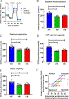
Orai inhibition reduces metabolic activity in CFS. A) Oxygen consumption rate (OCR) in CFS treated with Orai1 blockers CM-4620 (2 μM) or YM-58483 (2 μM), consecutively stimulated with oligomycin (oligo), FCCP, and antimycin/rotenone (AA + rot). Mitochondrial parameters were extrapolated, quantified, and represented in B) basal respiration, C) Maximal respiration, D) ATP turnover capacity, and E) Spare capacity. F) The energy profile of cultured fibroblasts at basal mitochondrial respiration and stressed mitochondrial respiration is based on the OCR and ECAR and among treatments within the same parameter. Data expressed as mean ± SD, one-way ANOVA (*p < 0.05), from three experiments from different cell preparations. Measurements are normalized to the total protein.
Figure 4.
Figure 4.
Orai activity is essential for maintaining mitochondrial metabolic flexibility in CFS. A-B) Basal OCR was established before injecting ETO followed by UK5099 and BPTES in untreated (Ctrl) (A), and CM4620 (2 μM) treated cells (B). C) % mitochondrial dependency, capacity, and flexibility percentage for oxidation of fatty acids calculated from A and B. D-E) Mitochondrial flexibility for glucose oxidation without (D) and with CM4620 treatment (E). Basal OCR was established before the injection of UK5099 and the following ETO and BPTES injection. F) % mitochondrial dependency and flexibility for glucose oxidation calculated from D and E. G-H) Mitochondrial flexibility for glutamine oxidation without (G) or with CM4620 treatment (H). Basal OCR was established before the sequential injection of BPTES and the following ETO and UK5099. I) % mitochondrial dependency and flexibility for glutamine oxidation calculated from G and H. The averages were taken after each injection. Data from three independent measurements are mean ± SD, n = 12 wells per group. Statistical differences were determined using an unpaired t-test (*p < 0.05). Abbreviations: fatty acids (FA), glucose (Glc), glutamine (Gln).
Figure 5.
Figure 5.
Orai inhibition alters cytosolic spatiotemporal ATP dynamics in CFS. A) Representative time course of cytosolic ATP dynamics in the presence of metabolic inhibitors, deoxyglucose (2DG), and oligomycin. B) Quantitation of cytosolic ATP content from A. C) Representative time course from CFS treated with CM4620 (1 h) and the addition of metabolic inhibitors. D) Quantitation of cytosolic ATP content from C. E) effect of Orai inhibition on cellular glucose uptake. F) effect of Orai inhibition on lactate production. Data are mean ± SD of triplicate samples obtained from a single experiment representative of at least three separate experiments. *P < 0.05, # P >0.05.
Figure 6.
Figure 6.
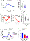
Orai channel is essential to maintain the ER Ca2+ and [Ca2+]mit homeostasis. A) Representative traces from MEFs showing thapsigargin-induced Ca2+ release from the ER store. The Ca2+ concentration was determined in fura-2-loaded cells exposed to 90-minute exposure to the Orai blocker (CM4620, blue trace). B) Quantitation of ER-Ca2+ content showing the average response to thapsigargin-induced Ca2+ release from the ER. Error bars are mean ± SD, (∗P < 0.05, n = 6). C) Representative traces from CFS expressing G-cepia1 er showing caffeine-induced ER Ca2+ discharge and refilling. D) Inhibition of Orai prevented Ca2+ re-entering the cell and refilling the ER. E) Absolute ER [Ca2+] was estimated after normalizing the fluorescence intensity change by dividing the baseline fluorescence before treatment. F) Mitochondrial Ca2+ uptake kinetics in MEF cells stimulated with ATP. Cells were perfused with a Ca2+-free KRB supplemented with 0.5 mM EGTA, in the presence of Orai inhibitors as indicated and finally stimulated with ATP (100 μM). G) Data are calculated as [Ca2+]mit peaks and presented as mean ± SEM, n = 6–8. *P<0.05, **P<00.01.

Similar articles

References

    1. Ebrahimighaei R, Tarassova N, Bond SC, McNeill MC, Hathway T, Vohra H, Newby AC, and Bond M. Extracellular matrix stiffness controls cardiac fibroblast proliferation via the nuclear factor-Y (NF-Y) transcription factor. Biochim Biophys Acta Mol Cell Res 1871: 119640, 2024. - PubMed
    1. Berridge MJ, Bootman MD, and Roderick HL. Calcium signalling: dynamics, homeostasis and remodelling. Nat Rev Mol Cell Biol 4: 517–529, 2003. - PubMed
    1. Crabtree GR. Calcium, calcineurin, and the control of transcription. The Journal of biological chemistry 276: 2313–2316, 2001. - PubMed
    1. Davis J, Burr AR, Davis GF, Birnbaumer L, and Molkentin JD. A TRPC6-dependent pathway for myofibroblast transdifferentiation and wound healing in vivo. Developmental cell 23: 705–715, 2012. - PMC - PubMed
    1. Feng J, Armillei MK, Yu AS, Liang BT, Runnels LW, and Yue L. Ca(2+) Signaling in Cardiac Fibroblasts and Fibrosis-Associated Heart Diseases. J Cardiovasc Dev Dis 6, 2019. - PMC - PubMed

LinkOut - more resources