

[18F]FDG PET imaging in the differentiation of cardiac masses: an updated systematic review and dual Meta-Analysis of diagnostic performance and parameter variability
- PMID: 40298987
- PMCID: PMC12222356
- DOI: 10.1007/s00259-025-07289-w
[18F]FDG PET imaging in the differentiation of cardiac masses: an updated systematic review and dual Meta-Analysis of diagnostic performance and parameter variability
Abstract
Introduction: Cardiac masses (CMs) encompass a heterogeneous group of benign, malignant, and pseudotumoural lesions, posing diagnostic challenges due to their rarity and varied aetiologies. Given the limitations of conventional imaging modalities in differentiating benign from malignant masses, [18F]FDG PET/CT has emerged as a promising technique by providing metabolic information. This systematic review and meta-analysis aimed to evaluate the diagnostic performance of [18F]FDG PET/CT in characterising CMs and assess semi-quantitative parameters' role in distinguishing malignant from benign lesions.
Methods: A systematic review and meta-analysis were conducted, including studies evaluating the diagnostic accuracy of [18F]FDG PET/CT in CMs. Sensitivity and specificity were pooled using a random-effects model, and a secondary analysis examined differences in SUVmax between malignant and benign lesions.
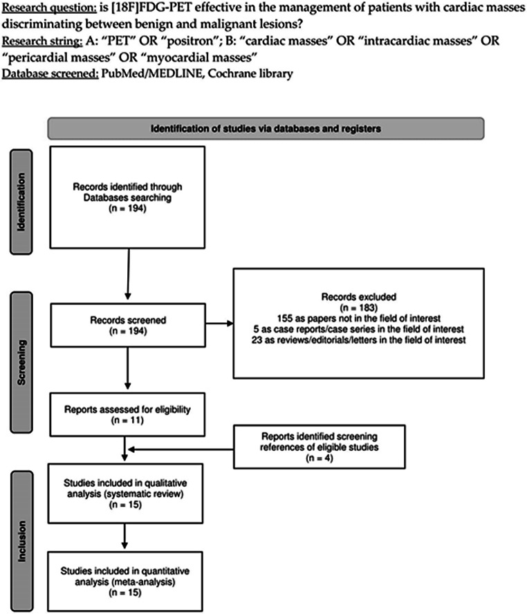
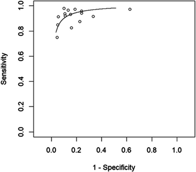
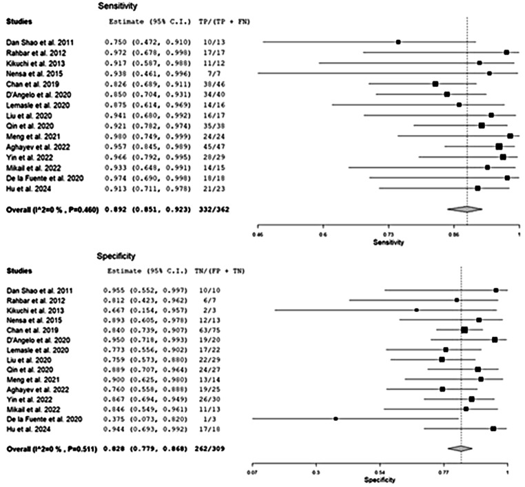
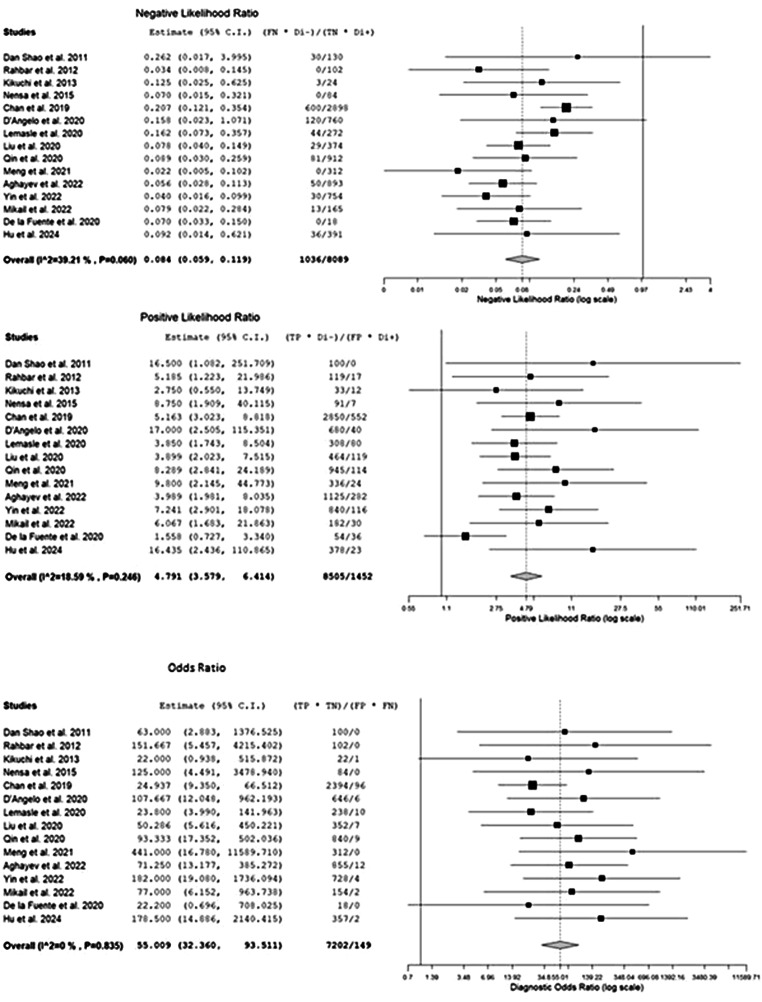
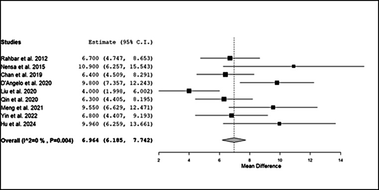
Results: Fifteen studies enrolling 1114 patients met inclusion criteria. The pooled sensitivity and specificity of [18F]FDG PET/CT in detecting malignant CMs were 89.2% (95% CI: 85-92%) and 82.8% (95% CI: 78-87%), respectively. Malignant lesions exhibited significantly higher SUVmax values (range: 5.6-14.3) than benign masses (range: 1.1-5.3, p < 0.001). PET/CT proved particularly effective in cases with inconclusive findings from echocardiography, cardiac magnetic resonance, or CT, contributing to biopsy guidance and treatment planning.
Conclusions: [18F]FDG PET/CT demonstrates robust diagnostic accuracy in differentiating benign from malignant cardiac masses, with SUVmax as a valuable malignancy marker. Its integration into multimodal imaging strategies enhances diagnostic certainty and optimises patient management. Despite these advantages, standardised imaging protocols and further multicentre prospective studies are warranted to refine its clinical application and validate its prognostic potential.
Keywords: Cardiac masses; Diagnostic accuracy; Meta-analysis; Oncology; [18F]FDG PET/CT.
© 2025. The Author(s).
Conflict of interest statement
Declarations. Ethical approval: Ethical approval was not required as this study was a systematic review and meta-analysis. Consent to participate: Informed consent was not necessary as this study did not include any human experiments. Consent to publish: The study does not contain any individual person’s data in any form (including any individual details, images or videos). Conflict of interest: The authors have no relevant financial or non-financial interests to disclose.
Figures
Similar articles
-
The value of FDG positron emission tomography/computerised tomography (PET/CT) in pre-operative staging of colorectal cancer: a systematic review and economic evaluation.Health Technol Assess. 2011 Sep;15(35):1-192, iii-iv. doi: 10.3310/hta15350. Health Technol Assess. 2011. PMID: 21958472 Free PMC article.
-
Fluorine-18-fluorodeoxyglucose (FDG) positron emission tomography (PET) computed tomography (CT) for the detection of bone, lung, and lymph node metastases in rhabdomyosarcoma.Cochrane Database Syst Rev. 2021 Nov 9;11(11):CD012325. doi: 10.1002/14651858.CD012325.pub2. Cochrane Database Syst Rev. 2021. PMID: 34753195 Free PMC article.
-
Imaging modalities for characterising focal pancreatic lesions.Cochrane Database Syst Rev. 2017 Apr 17;4(4):CD010213. doi: 10.1002/14651858.CD010213.pub2. Cochrane Database Syst Rev. 2017. PMID: 28415140 Free PMC article.
-
Whole-Body 18F-FDG PET and 18F-FDG PET/CT in Patients with Suspected Paraneoplastic Syndrome: A Systematic Review and Meta-Analysis of Diagnostic Accuracy.J Nucl Med. 2017 Jul;58(7):1031-1036. doi: 10.2967/jnumed.116.183905. Epub 2016 Dec 15. J Nucl Med. 2017. PMID: 27980049
-
Prognostic value of 18F-FDG-PET/CT parameters in patients with pancreatic carcinoma: A systematic review and meta-analysis.Medicine (Baltimore). 2017 Aug;96(33):e7813. doi: 10.1097/MD.0000000000007813. Medicine (Baltimore). 2017. PMID: 28816978 Free PMC article.
Cited by
-
A Comprehensive Review of Cardiac Tumors: Imaging, Pathology, Treatment, and Challenges in the Third Millennium.Diagnostics (Basel). 2025 May 30;15(11):1390. doi: 10.3390/diagnostics15111390. Diagnostics (Basel). 2025. PMID: 40506961 Free PMC article. Review.
References
-
- Angeli F, Fabrizio M, Paolisso P, Magnani I, Bergamaschi L, Bartoli L, et al. [Cardiac masses: classification, clinical features and diagnostic approach]. G Ital Cardiol (Rome). 2022;23:620–30. - PubMed
-
- Jain S, Dhingra V, Girdhani B. Scope of PET imaging in the evaluation of cardiac tumors. Cancer Treat Res Commun. 2023;37:100754. - PubMed
-
- Burke A, Tavora F. The 2015 WHO classification of tumors of the heart and pericardium. J Thorac Oncol. 2016;11:441–52. - PubMed
Publication types
MeSH terms
Substances
LinkOut - more resources
Full Text Sources
Miscellaneous
