. 2025 Apr 3;13(4):872.
doi: 10.3390/biomedicines13040872.

TGF-β1 Improves Nerve Regeneration and Functional Recovery After Sciatic Nerve Injury by Alleviating Inflammation

Affiliations

TGF-β1 Improves Nerve Regeneration and Functional Recovery After Sciatic Nerve Injury by Alleviating Inflammation

Maorong Jiang et al. Biomedicines. .

Abstract

Background: Peripheral nerves have a certain regenerative ability, but their repair and regeneration after injury is a complex process, usually involving a large number of genes and proteins. In a previous study, we analyzed the gene expression profile in rats after sciatic nerve injury and found significant changes in transforming growth factor-beta 1 (TGF-β1) expression, suggesting that TGF-β1 may be involved in the process of nerve regeneration after injury. Methods: In this study, we first detected the time-course expression and localization of TGF-β1 in dorsal root ganglion (DRG) tissues in a rat sciatic nerve transection model via RT-qPCR. Secondly, we investigated the bioactive roles of TGF-β1 in primary cultured DRG neuron cells through a CCK8 assay, TUNEL assay, and immunofluorescence staining. Thirdly, we explored the neuroprotective roles of TGF-β1 in an in vivo model of sciatic nerve regeneration through morphological observation, behavioral, and electrophysiological tests, and a molecular biological measure. Results: We found that TGF-β1 expression was increased after injury and mainly located in the cytoplasm and nuclei of neuron cells in the DRG. TGF-β1 may regulate the viability, apoptosis, and neurite outgrowth of primary DRG neuron cells. In our in vivo model of sciatic nerve regeneration, TGF-β1 improved nerve regeneration and neuronal function recovery after sciatic nerve injury, alleviated the inflammatory response, and relieved neuropathic pain via the TGF-β1/smad2 pathway. Conclusions: This study provides an experimental and theoretical basis for using TGF-β1 as a neuroprotective agent after peripheral nerve injury in clinical practice in the future.

Keywords: TGF-β1; inflammatory response; nerve regeneration; neuronal function recovery; peripheral nerve injury.


Conflict of interest statement

The authors declare that they have no competing interests.

Figures

Figure 1
Figure 1
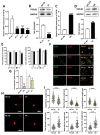
Time-course expression and localization of TGF-β1. (A,B) Time-course mRNA expression of TGF-β1 in L4 and L5 DRG tissues in rats after sciatic nerve transection via RT-qPCR. Versus od, * p < 0.05 and ** p < 0.01. (C) Expression and localization of TGF-β1 in DRG tissue via immunofluorescence staining. The right image is an enlarged version of the box on the left. Scale bar = 100 μm. (D) Expression and localization of TGF-β1 in DRG neuron cells via immunocytochemistry staining. The primary DRG neuron cells were cultured by digesting DRG tissues. Scale bar = 50 μm. n = 3. TGF-β1, transforming growth factor-beta 1. RT-qPCR, real-time quantitative PCR. DRG, dorsal root ganglia.
Figure 2
Figure 2
Effects of TGF-β1 on DRG neurons. The shRNA lentivirus (sh-a, sh-b, and sh-c) was prepared for the knockdown of TGF-β1, and lentivirus (OE) was prepared for the overexpression of TGF-β1. (A) The expressions of TGF-β1 in DRG neuron cells after transfection with shRNA lentivirus were detected via RT-qPCR. (B) The expressions of TGF-β1 in DRG neuron cells after transfection with shRNA lentivirus (sh-c) were detected via Western blot. Versus NC-sh, ** p < 0.01 and *** p < 0.001. (C,D) The expressions of TGF-β1 in DRG neuron cells after transfection with overexpression lentivirus were detected via RT-qPCR or Western blot. Versus NC-OE, ** p < 0.01 and *** p < 0.001. (E) Cell viability of DRG neurons after transfection with shRNA lentivirus or overexpression lentivirus prior to 1 d, 3 d, or 5 d. Versus NC-sh or NC-OE, * p < 0.05 and ** p < 0.01. (F) TUNEL staining of DRG neuron cells after transfection with shRNA lentivirus or overexpression lentivirus. Scale bar = 100 μm. (G) Statistical analysis of cell apoptosis (F). Versus NC-sh or NC-OE, * p < 0.05 and ** p < 0.01. (H) Immunofluorescence staining with Tuj1 of DRG neuron cells after transfection with shRNA lentivirus or overexpression lentivirus. (I) Statistical analysis of total neurite length, mean neurite length, and longest neurite length of DRG neurons. *** p < 0.001. TGF-β1, transforming growth factor-beta 1. RT-qPCR, real-time quantitative PCR. DRG, dorsal root ganglia. shRNA, small hairpin RNA. NC-sh, shRNA lentivirus negative control. OE, overexpression. NC-OE, overexpression lentivirus negative control. TUNEL, TdT-mediated dUTP Nick-End Labeling.
Figure 3
Figure 3
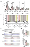
Effects of TGF-β1 on cell factors in DRG neurons. (A) The mRNA expressions of β-catenin, Akt, smad2, Nf-κB, IL-10, TNF-α, and bFGF in DRG neuron cells after transfection with shRNA lentivirus or overexpression lentivirus were detected via RT-qPCR. (B) The expressions of β-catenin, Akt, p-Akt, smad2, p-smad2, and Nf-κB in DRG neuron cells after transfection with shRNA lentivirus or overexpression lentivirus were detected via Western blot. (C) Statistical analysis of (B). Versus NC-sh or NC-OE, * p < 0.05, ** p < 0.01 and *** p < 0.001. TGF-β1, transforming growth factor-beta 1. RT-qPCR, real-time quantitative PCR. DRG, dorsal root ganglia. shRNA, small hairpin RNA. NC-sh, shRNA lentivirus negative control. OE, overexpression. NC-OE, overexpression lentivirus negative control. Nf-κB, nuclear factor kappa-B. IL, interleukin. TNF-α, tumor necrosis factor-α. bFGF, basic fibroblast growth factor. GAPDH, glyceraldehyde-3-phosphate dehydrogenase.
Figure 4
Figure 4
Expressions of TGF-β1 in in vivo model after lentivirus injection. After the rat sciatic nerve regeneration model was established, the lentivirus was intrathecally injected into the L4-L5 foramina. (A) The mRNA expressions of TGF-β1 in the DRG at 7 days or 10 days after injection with overexpression lentivirus were detected via RT-qPCR. (B) The expressions of TGF-β1 in the DRG at 10 days after injection with overexpression lentivirus were detected via Western blot. (C) The mRNA expressions of TGF-β1 in the DRG at 3 days after injection with shRNA lentivirus were detected via RT-qPCR. (D) The expressions of TGF-β1 in the DRG at 3 days after injection with shRNA lentivirus (sh-c) were detected via Western blot. (E) Immunofluorescence staining with Tuj1 in the DRG after transfection with shRNA lentivirus. The recombination virus vector included the GFP gene. Scale bar = 50 μm. Versus NC-sh or NC-OE, * p < 0.05, and ** p < 0.01. n = 3. TGF-β1, transforming growth factor-beta 1. RT-qPCR, real-time quantitative PCR. DRG, dorsal root ganglia. shRNA, small hairpin RNA. NC-sh, shRNA lentivirus negative control. OE, overexpression. NC-OE, overexpression lentivirus negative control. GFP, green fluorescent protein.
Figure 5
Figure 5
Effects of TGF-β1 on functional recovery after sciatic nerve injury. After the rat sciatic nerve regeneration model was established, the lentivirus was intrathecally injected into the L4-L5 foramina. The thermal pain and sciatic nerve function index were measured at 1, 2, 4, 6, and 8 weeks after lentivirus injection. (A) The statistical analysis of withdrawal reflex latency. (B) The statistical analysis of sciatic nerve functional index. The compound muscle action potentials (CMAPs) were recorded at 8 weeks after virus injection. (C) The representative images of CMAPs. The statistical analysis of latency (D) and CMAP amplitude (E). * p < 0.05, and ** p < 0.01. n = 3. TGF-β1, transforming growth factor-beta 1. shRNA, small hairpin RNA. NC-sh, shRNA lentivirus negative control. OE, overexpression. NC-OE, overexpression lentivirus negative control.
Figure 6
Figure 6
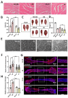
Effects of TGF-β1 on nerve regeneration after sciatic nerve injury. After the rat sciatic nerve regeneration model was established, the lentivirus was intrathecally injected into the L4-L5 foramina. The H&E staining of gastrocnemius muscles, immunofluorescence staining of SCG10, and transmission electron microscope examination of sciatic nerve were performed at 8 weeks after virus injection. (A) Representative images of H&E staining. (B) Statistical analysis of cross-sectional area of muscle fibers. (C) Representative images of gastrocnemius muscles. The left gastrocnemius muscles were from operated limbs, the right muscles were from contralateral limbs. (D) Statistical analysis of wet weight ratio of gastrocnemius muscles. (E) Representative images of transmission electron microscopy. Scale bar = 5 μm. (F) Statistical analysis of myelin sheath thickness. (G) Immunofluorescence staining of SCG10 of sciatic nerve. Scale bar = 200 μm. (H) Statistical analysis of relative fluorescence intensity. * p < 0.05 and ** p < 0.01. n = 3. H&E, Haematoxylin and Eosin. TGF-β1, transforming growth factor-beta 1. shRNA, small hairpin RNA. NC-sh, shRNA lentivirus negative control. OE, overexpression. NC-OE, overexpression lentivirus negative control.
Figure 7
Figure 7
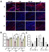
Effects of TGF-β1 on inflammation after sciatic nerve injury. (A) The immunofluorescence staining of F4/80 in the sciatic nerve was performed 1, 4, and 7 days after lentivirus injection. (B) Statistical analysis of relative fluorescence intensity. (C) The mRNA expressions of TNF-α and IL-10 in the sciatic nerve were detected via RT-qPCR. Versus NC-sh or NC-OE, * p < 0.05, and ** p < 0.01. n = 3. TGF-β1, transforming growth factor-beta 1. RT-qPCR, real-time quantitative PCR. shRNA, small hairpin RNA. NC-sh, shRNA lentivirus negative control. OE, overexpression. NC-OE, overexpression lentivirus negative control. IL, interleukin. TNF-α, tumor necrosis factor-α.
Figure 8
Figure 8
Effects of TGF-β1 on cell factors after sciatic nerve injury. After the rat model of sciatic nerve regeneration was established, the lentivirus was intrathecally injected into the L4-L5 foramina. (A) The mRNA expressions of β-catenin, Akt, smad2, Nf-κB, IL-10, TNF-α, NP2, and bFGF in the DRG after lentivirus injection were detected via RT-qPCR. (B) The expressions of β-catenin, Akt, p-Akt, smad2, p-smad2, and Nf-κB in the DRG after lentivirus injection were detected via Western blot. (C) Statistical analysis of (B). Versus NC-sh or NC-OE, * p < 0.05, and ** p < 0.01. n = 3. TGF-β1, transforming growth factor-beta 1. RT-qPCR, real-time quantitative PCR. DRG, dorsal root ganglia. shRNA, small hairpin RNA. NC-sh, shRNA lentivirus negative control. OE, overexpression. NC-OE, overexpression lentivirus negative control. Nf-κB, nuclear factor kappa-B. IL, interleukin. TNF-α, tumor necrosis factor-α. NP2, neuronal pentraxin 2. bFGF, basic fibroblast growth factor. GAPDH, glyceraldehyde-3-phosphate dehydrogenase.

Similar articles

Cited by

References

    1. Juckett L., Saffari T.M., Ormseth B., Senger J.-L., Moore A.M. The Effect of Electrical Stimulation on Nerve Regeneration Following Peripheral Nerve Injury. Biomolecules. 2022;12:1856. doi: 10.3390/biom12121856. - DOI - PMC - PubMed
    1. Divac N., Aksić M., Rasulić L., Jakovčevski M., Basailović M., Jakovčevski I. Pharmacology of repair after peripheral nerve injury. Int. J. Clin. Pharmacol. Ther. 2021;59:447–462. doi: 10.5414/CP203893. - DOI - PubMed
    1. Contreras E., Bolívar S., Navarro X., Udina E. New insights into peripheral nerve regeneration: The role of secretomes. Exp. Neurol. 2022;354:114069. doi: 10.1016/j.expneurol.2022.114069. - DOI - PubMed
    1. O’brien A.L., West J.M., Saffari T.M., Nguyen M., Moore A.M. Promoting Nerve Regeneration: Electrical Stimulation, Gene Therapy, and Beyond. Physiology. 2022;37:302–310. doi: 10.1152/physiol.00008.2022. - DOI - PubMed
    1. Carvalho C.R., Reis R.L., Oliveira J.M. Fundamentals and Current Strategies for Peripheral Nerve Repair and Regeneration. Adv. Exp. Med. Biol. 2020;1249:173–201. - PubMed

LinkOut - more resources