. 2025 Apr 14;13(4):958.
doi: 10.3390/biomedicines13040958.

Ivosidenib Confers BRCAness Phenotype and Synthetic Lethality to Poly (ADP-Ribose) Polymerase Inhibition in BRCA1/2-Proficient Cancer Cells

Affiliations

Ivosidenib Confers BRCAness Phenotype and Synthetic Lethality to Poly (ADP-Ribose) Polymerase Inhibition in BRCA1/2-Proficient Cancer Cells

Danyang Zhou et al. Biomedicines. .

Abstract

Background/Objectives: PARP inhibitors (PARPi) are pivotal to treating homologous recombination repair-deficient (HRD) cancers, particularly BRCA1/2-mutated ovarian and breast cancers. However, most ovarian and breast cancers harbor wild-type (WT) BRCA1/2, limiting PARPi eligibility. This study aims to identify an approved drug that could induce a BRCAness phenotype, thereby sensitizing WT BRCA cancers to PARPi. Methods: Ovarian and breast cancer cell lines with WT BRCA1/2 were treated with ivosidenib. HR repair efficiency was assessed via RAD51 foci formation and reporter assays. Synthetic lethality with PARPi was evaluated using viability and colony formation assays. Mechanistic studies included RNA-binding protein pulldown, co-immunoprecipitation, and functional analyses of DNA repair pathways. YTHDC2's role in HR was investigated through siRNA knockdown and rescue experiments. Results: Ivosidenib significantly reduced HR repair efficiency and sensitized cells to PARPi, inducing synthetic lethality. Mechanistically, ivosidenib directly bound YTHDC2, an m6A reader critical for HR. This interaction disrupted YTHDC2's ability to promote DNA double-strand break repair via HR, evidenced by impaired recruitment of repair proteins (e.g., BRCA1, RAD51) and accumulation of DNA damage (γH2AX foci). YTHDC2 knockdown phenocopied ivosidenib effects, while overexpression rescued HR defects. Conclusions: Ivosidenib induces BRCAness in WT BRCA ovarian and breast cancers by targeting YTHDC2, thereby suppressing HR repair and enhancing PARPi sensitivity. This uncovers a novel, metabolism-independent mechanism of ivosidenib, repositioning it as a therapeutic agent for HRD tumors. These findings propose a strategy to expand PARPi eligibility to WT BRCA cancers, addressing a critical unmet need in oncology.

Keywords: BRCA1/2; HR repair; Ivosidenib; PARP inhibitors; ovarian cancer.


Conflict of interest statement

The authors declare no conflicts of interest.

Figures
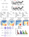
Figure 1
Figure 1
High-throughput screening shows that IDH1 inhibitor ivosidenib greatly sensitizes BRCA1/2-proficient cancer cells to PARPi. (A) The workflow for screening sensitizer drugs for the PARP inhibitor Olaparib in A2780 cells using an FDA-approved drug library (TargetMol, #L1000) that contains 2040 approved small molecule drugs. (B) The relative cell viability of A2780 cells grown in the presence of 10 μM of FDA-approved drugs alone (upper) or FDA-approved drugs plus 10 μM of Olaparib for 72 h; cell viability was analyzed through CCK-8 assay. (C) Ivosidenib increases Olaparib sensitivity in BRCA1/2-WT breast and ovarian cancer cells. Indicated cells were treated with increasing concentrations of Olaparib (ola) in the presence or absence of 5 μM of ivosidenib (Ivo) for 72 h, followed by cell viability analysis. (D) Ivosidenib and Olaparib synergistically suppress cancer cell growth. Indicated cells were co-treated with serial dosages of Olaparib and ivosidenib for 72 h, followed by Bliss synergy analysis. (E) Ivosidenib and Olaparib synergistically inhibit clonogenic survival in BRCA1/2-WT cancer cells. The colony formation analysis indicated cells treated with 2 μM of ivosidenib, 2 μM of Olaparib, or their combination. Data are presented as means ± SD, n = 6. * p < 0.05 and *** p < 0.001 based on the two-tailed t-test.
Figure 2
Figure 2
Ivosidenib (Ivo) greatly potentiates Olaparib (Ola)-induced DNA damage in BRCA1/2-proficient cells. (A) Ivosidenib enhances Olaparib-induced γH2AX formation in BRCA1/2-WT cancer cells. Indicated cells were treated with 50 μM of Ola, 10 μM of Ivo, or their combination for 48 h, followed by Western blot analysis of γH2AX. (B) Ivosidenib delayed the reduction of γH2AX after Olaparib treatment. A2780 cells were treated with 100 μM for 48 h and then released into normal culture medium in the presence or absence of Ivo for indicated time points, followed by Western blot analysis of γH2AX. (C) Ivosidenib increases Olaparib-caused DNA damage. A2780 cells were treated with 50 μM of Ola, 10 μM of Ivo, or their combination for 48 h, followed by measuring the DNA damage through comet assay. Data are presented as means ± SD.
Figure 3
Figure 3
Ivosidenib (Ivo) treatment suppresses the repair of Olaparib (Ola)-induced DSBs. Ivosidenib inhibits HR repair. (A,B) Ivosidenib A2780 cells were treated with 50 of μM Ola, 10 μM of Ivo, or their combination for 48 h, followed by immunofluorescence analysis of Rad51/γH2AX (A) or RPA2/γH2AX (B). (C) Cells were treated as indicated for 48 h, followed by Western blot analysis of pRPA2 S4/8. Data were are presented as means ± SD. ** p < 0.01 and *** p < 0.001 according to the two2-tailed t-test.
Figure 4
Figure 4
Ivosidenib targets YTHDC2. (AC) Ivosidenib protects pronase-mediated digestion of YTHDC2. (A) 200 μg of A2780 cell lysates was incubated with/without 10 μM of ivosidenib (Ivo) before being subjected to pronase digestion; the protein contents were analyzed through Coomassie staining and mass spectrometry analysis. * indicates the protein YTHDC2. The drug affinity responsive target stability (DARTS) detection of single-dose (B) or serial-dosage (C) Ivo prevention of pronase-mediated YTHDC2 digestion. (D) Molecular docking shows the binding pocket of Ivo in YTHDC2 protein.
Figure 5
Figure 5
YTHDC2 knockdown suppresses HR repair. (A) Schematic diagram of HR repair reporter system (DR-GFP). (B,C) Silencing of YTHDC2 decreases HR repair. QU20S-DRGFP cells were transfected with control (ctrl) or YTHDC2 siRNA and, 24 h later, cells were transfected with pCBASceI plasmid to induce endonuclease-induced DSB, followed by flow cytometry analysis of GFP expression 48 h after pCBASceI expression (B). A2780 cells were transfected with control (Ctrl) or YTHDC2 siRNA; cells were then treated with 50 μM of Olaparib for 48 h, followed by immunofluorescence analysis of Rad51/γH2AX (C). Data are presented as means ± SD. *** p < 0.001 according to the two-tailed t-test.
Figure 6
Figure 6
Depletion of YTHDC2 abolished ivosidenib-induced reduction of Rad51 foci formation. (A) YTHDC2 was depleted in A2780 cells using the CRISPR/Cas9 technique. (B,C) Ivosidenib inhibits Rad51 foci by targeting YTHDC2. WT or YTHDC2 knockout A2780 cells were pre-treated with or without 10 μM of ivosidenib (Ivo) for 2 h; then, cells were irradiated with 2 Gy X-ray, and Rad51/γH2AX foci formation was then analyzed. The representative Rad51/γH2AX staining (B) and quantification of Rad51 positive cells (C) were shown. Data are presented as means ± SD. ** p < 0.01 according to the two-tailed t-test. n.s., no significance.
Figure 7
Figure 7
Combination treatment of ivosidenib (Ivo) and Olaparib (Ola) produces synergistic anti-tumor efficacy in BRCA1/2-proficient ovarian cancers. (AC) Ivosidenib and Olaparib synergistically suppress tumor growth. A2780 xenograft bearing mice treated with Ola (100 mg/kg, orally) and Ivo (100 mg/kg, orally) and tumor growth curve (A), tumor image (B), and tumor weights (C) are shown. (D,E) Co-treatment with ivosidenib and Olaparib greatly induces DSBs and reduces proliferation in A2780 xenograft tumors. Immunohistochemistry analysis of γH2AX (D) and Ki67 (E) in tumors treated as indicated. Data are presented as means ± SD. *** p < 0.001 according to the two-tailed t-test.

Similar articles

References

    1. Chappidi N., Quail T., Doll S., Vogel L.T., Aleksandrov R., Felekyan S., Kuhnemuth R., Stoynov S., Seidel C.A.M., Brugues J., et al. PARP1-DNA co-condensation drives DNA repair site assembly to prevent disjunction of broken DNA ends. Cell. 2024;187:945–961.e918. doi: 10.1016/j.cell.2024.01.015. - DOI - PubMed
    1. Jackson S.P., Bartek J. The DNA-damage response in human biology and disease. Nature. 2009;461:1071–1078. doi: 10.1038/nature08467. - DOI - PMC - PubMed
    1. Chatterjee N., Walker G.C. Mechanisms of DNA damage, repair, and mutagenesis. Environ. Mol. Mutagen. 2017;58:235–263. doi: 10.1002/em.22087. - DOI - PMC - PubMed
    1. Alemasova E.E., Lavrik O.I. Poly(ADP-ribosyl)ation by PARP1: Reaction mechanism and regulatory proteins. Nucleic Acids Res. 2019;47:3811–3827. doi: 10.1093/nar/gkz120. - DOI - PMC - PubMed
    1. Herrmann G.K., Yin Y.W. The Role of Poly(ADP-ribose) Polymerase 1 in Nuclear and Mitochondrial Base Excision Repair. Biomolecules. 2023;13:1195. doi: 10.3390/biom13081195. - DOI - PMC - PubMed

LinkOut - more resources