. 2025 Apr 29;10(11):e177373.
doi: 10.1172/jci.insight.177373. eCollection 2025 Jun 9.

Identification and regulation of circulating tumor-TCR-matched cytotoxic CD4+ lymphocytes by KLRG1 in bladder cancer

Affiliations

Identification and regulation of circulating tumor-TCR-matched cytotoxic CD4+ lymphocytes by KLRG1 in bladder cancer

Serena S Kwek et al. JCI Insight. .

Abstract

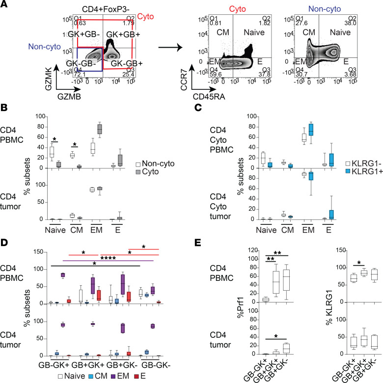
While cytotoxic CD4+ tumor-infiltrating lymphocytes have anticancer activity in patients, whether these can be noninvasively monitored and how these are regulated remains obscure. By matching single cells with T cell receptors (TCRs) in tumor and blood of patients with bladder cancer, we identified distinct pools of tumor-matching cytotoxic CD4+ T cells in the periphery directly reflecting the predominant antigenic specificities of intratumoral CD4+ tumor-infiltrating lymphocytes. On one hand, the granzyme B-expressing (GZMB-expressing) cytotoxic CD4+ subset proliferated in blood in response to PD-1 blockade but was separately regulated by the killer cell lectin-like receptor G1 (KLRG1), which inhibited their killing by interacting with E-cadherin. Conversely, a clonally related, GZMK-expressing circulating CD4+ population demonstrated basal proliferation and a memory phenotype that may result from activation of GZMB+ cells, but was not directly mobilized by PD-1 blockade. As KLRG1 marked the majority of circulating tumor-TCR-matched cytotoxic CD4+ T cells, this work nominates KLRG1 as a means to isolate them from blood and provide a window into intratumoral CD4+ recognition, as well as a putative regulatory receptor to mobilize the cytolytic GZMB+ subset for therapeutic benefit. Our findings also underscore ontogenic relationships of GZMB- and GZMK-expressing populations and the distinct cues that regulate their activity.

Keywords: Cancer immunotherapy; Cellular immune response; Clinical trials; Immunology; Oncology; T cells.


Conflict of interest statement

Conflict of interest: DYO has received research support from Merck, PACT Pharma, Parker Institute for Cancer Immunotherapy, Poseida Therapeutics, TCR2 Therapeutics, Roche/Genentech, Nutcracker Therapeutics, Amgen, and Allogene Therapeutics; travel and accommodations from Roche/Genentech, Poseida Therapeutics, and DAVA Oncology; and has consulted for Revelation Partners. LF has received research support from Roche/Genentech, Abbvie, Bavarian Nordic, Bristol Myers Squibb, Dendreon, Janssen, Merck, and Partner Therapeutics; and has served on scientific advisory boards for Actym, AstraZeneca, Atreca, Bioatla, Bolt, Bristol Myers Squibb, Daiichi Sankyo, Immunogenesis, Innovent, Merck, Merck KGA, Nutcracker, RAPT, Scribe, Senti, Sutro, and Roche/Genentech. EDC and CJY are co-founders of Survey Genomics. CJY is co-founder of Dropprint Genomics.

Figures

Figure 1
Figure 1. Combined blood and tumor TCR repertoire network analysis.
(A) Blood and tumor samples were obtained from standard-of-care (SOC) and anti–PD-L1–treated patients with bladder cancer (on trial NCT02451423) for scRNA-seq/scTCR-seq, using matching TCRs on single cells as a barcode for matched antigenic specificity between compartments. (B) UMAP plot of 157,054 single cells clustered together, where each phenotypic population is identified with a distinct color and density plots showing distribution of cells for the subset of samples sorted for CD8+ or CD4+ T cells, or obtained from normal adjacent tissue (NAT), tumor, and blood. n = 7 tumors, 6 NAT, and 7 matched PBMC samples (before and after treatment for immunotherapy-treated patients). (C) Dot plot showing fractions of cells and mean expression of selected genes (bottom labels) in each cell cluster (side labels). (D) Feature plots showing expression of transcripts GZMB, GZMK, or KI67 (red) in the cells from samples sorted for CD4+ cells (gray) from blood or tumor superimposed on the UMAP plot. CD4+ cells expressing cytotoxic transcripts are known to express CD4 protein based on sorting, but are coclustered with a predominance of CD8+ T cells due to their cytotoxic gene expression. (E) Phenotypic analysis of all CD4+ T cells using gene signatures for Th1, Th2, and Trm (pairwise comparison statistics in Supplemental Dataset 5). (F) Percentage of TCRs that are unique, expanded (cluster size >1), and expanded and blood-tumor matched for CD4+ and CD8+ T cells from blood and tumor. (G) Network TCR plots of a representative patient. Each dot represents 1 cell, and the dots within in each cluster in red have identical paired TCRαβ.
Figure 2
Figure 2. CD4+ and CD8+ T cells in PBMCs and tumor with matching TCRs are predominantly cytotoxic.
(A) Box-and-whisker plots showing percentages of cells in each phenotypic population. Proportion of each cell phenotype within all TCRs, expanded TCRs, and blood- and tumor-matched TCRs in CD4+ and CD8+ T cells from blood and tumor. Statistical results of pairwise comparison for each population with the other populations are shown in Supplemental Dataset 6. n = 6 paired tumors and PBMCs. (B) Box-and-whisker plots with whiskers (median and interquartile ranges) showing number of TCR clusters containing each cell phenotype within matched and nonmatched TCRs in the blood. *FDR < 0.05. Linear mixed-model statistics are shown in Supplemental Dataset 7. n = 6 paired tumors and PBMCs. (C) Volcano plots showing DE genes of cells with matched TCRs versus cells with nonmatched TCRs from CD4+ and CD8+ T cells in the blood. Top genes by FDR are shown. Red highlights positive coefficient and blue negative coefficient. DE statistics in Supplemental Dataset 8.
Figure 3
Figure 3. Phenotypic heterogeneity and plasticity of individual TCR clusters.
(A) A representative CD4+ T cell network cluster with matching TCR clonotype from blood and tumor, and a separate CD8+ T cell network cluster with a different matching TCR clonotype. Each dot is 1 cell and the color represents a different cellular phenotype. Tables show the number of cells with the indicated phenotype in the cluster. (B) Pseudotime trajectories of GZMB+, GZMK+, CXCL13+, Tregs, Prolif, and MAIT cells with blood- and tumor-matched TCRs in CD4+ and CD8+ T cells from 2 untreated standard-of-care (SOC) patients and 3 patients treated with anti–PD-L1 (atezolizumab, Atezo) on trial. Black circles are branch nodes. Major branches A, B, and C are labeled. (C) Plots showing percentage of Prolif cell phenotype in each TCR cluster from blood- and tumor-TCR-matched clusters in blood before and after anti–PD-L1 treatment. The number (n) of clusters is listed below the plots. *P < 0.05 by nonparametric Mann-Whitney U test. (D) Radar plots of mean percentage of selected cell phenotype in each TCR cluster in matched TCR clusters of pretreatment PBMCs (white circles), post-Atezo-treatment PBMCs (blue circles), and post-Atezo-treatment tumors (red circles) for CD4+ and CD8+ T cells.
Figure 4
Figure 4. Expression of proliferative and inhibitory markers on cytotoxic versus noncytotoxic cell types in PBMCs and bladder tumors.
(A) Normalized log(fold change) from scRNA-seq analysis for selected phenotypes and genes. (B) Flow cytometry was carried out on 8 PBMC samples and 6 tumors from standard-of-care (SOC) patients with bladder cancer. (C) Representative flow cytometry plots showing CD4+FoxP3 T cells expressing GZMB and GZMK, and total cytotoxic subtypes (Cyto) and noncytotoxic subtypes (Non-cyto) subtypes are gated as shown. (D) Box-and-whisker plots showing percentage Ki67+, PD-1+, Tim-3+, and KLRG1+ of Cyto and Non-Cyto from CD4+FoxP3 T cells. (E) Box-and-whisker plots showing percentage PD-1+, Tim-3+, and KLRG1+ of Ki67+ and Ki67cytotoxic cells. (F) Box-and-whisker plots showing percentage PD-1+ and Tim-3+ of KLRG1+ and KLRG1 cytotoxic cells. Comparison of paired cell subsets within PBMCs or tumors was performed using Friedman’s test with Dunn’s multiple-comparison test. Comparison of nonpaired cell subsets between PBMCs and tumors was performed using Kruskal-Wallis test with Dunn’s multiple-comparison test. *P < 0.05; **P < 0.01; ***P < 0.001. Asterisks in black, red, or blue indicate significant differences between subsets within PBMCs, within tumors, and between PBMCs and tumors, respectively. Corresponding plots for CD8+ cells are shown in Supplemental Figure 4.
Figure 5
Figure 5. Developmental stages of different cytotoxic cell types in PBMCs and bladder tumors.
(A) Flow cytometry was carried out on 8 PBMC samples and 6 tumors from standard-of-care patients with bladder cancer. Representative flow cytometry plots showing CD4+FoxP3 T cells expressing GZMB and GZMK, and Cyto and Non-Cyto cells were further gated into Naive, CM, EM, and E based on expression of CCR7 and CD45RA. All graphs in this figure are box-and-whisker plots for CD4+ T cells from PBMCs and tumors showing (B) proportion of developmental subsets in Cyto and Non-cyto cells; (C) proportion of developmental subsets in KLRG1 and KLRG1+ cytotoxic cells; (D) proportion of developmental subsets in each cytotoxic subtype (red, purple, and black lines indicate significant pairwise comparison of proportion of E, EM, or Naive subsets, respectively, between cytotoxic subtypes); and (E) percentage perforin+ (Prf1+) and KLRG1+ of each cytotoxic subtype. Comparison of paired cell subsets was performed using Friedman’s test with Dunn’s multiple-comparison test. *P < 0.05; **P < 0.01; ****P < 0.0001. Corresponding plots for CD8+ cells are shown in Supplemental Figure 4.
Figure 6
Figure 6. Anti–PD-L1 treatment increased Ki67 and decreased KLRG1 expression on cytotoxic T cells in the blood.
(A) Percentage of cells expressing either Mki67 or Klrg1 transcript in each TCR cluster from pre- and post-atezolizumab patient PBMCs. Each line represents a different TCR cluster. Friedman’s test with Dunn’s multiple-comparison test was carried out pre- and post-treatment timepoints for CD4+ (red) and CD8+ (black) TCR clusters (TCR clusters = 13). (B) Stacked bar plot showing mean percentage of cells expressing Mki67 and/or Klrg1 (KL) transcripts before and after anti–PD-L1 treatment, as shown per TCR cluster (TCR clusters = 13). (C) Flow cytometry was carried out on PBMCs from 8 healthy individuals and 14 pre- and post-atezolizumab-treated patients with bladder cancer. (D) Representative flow cytometry of pre- and posttreatment CD4+ EM cytotoxic T cells showing Ki67 and KLRG1 expression. (E) Percentage KLRG1Ki67+ among total CD4+ T cells in cytotoxic subtypes with Naive-like, CM, EM, and E phenotypes. (F) Comparisons of pretreatment percentage of KLRG1Ki67+ among total CD4+ T cells between cytotoxic subsets. Mann-Whitney U test was used to compare healthy and pretreatment samples; Wilcoxon’s matched-pairs signed-rank t test was used to compare pre- and posttreatment samples. *P < 0.05; **P < 0.01; ***P < 0.001. Corresponding plots for CD8+ cells are in Supplemental Figure 6.
Figure 7
Figure 7. Expression and function of KLRG1 in cytotoxic CD4+ T cells.
(A) Box-and-whisker plot showing percentage of KLRG1+Ki67 T cells among 14 post-atezolizumab-treatment EM and E cytotoxic subsets in PBMCs. Friedman’s test with Dunn’s multiple-comparison test was carried out. *P < 0.05; **P < 0.01; ****P < 0.0001. (B) Strategy for sorting and autologous tumor killing assay. (C) Flow cytometry of E-cadherin staining of live CD45CD3 cells in a portion of tumors used in the killing assay, and KLRG1+ and KLRG1 gates of CD4+(CD127loCD25+) cells from PBMCs and tumors sorted for T cell expansion. (D) Apoptotic tumor cell death plotted as the relative change in annexin V+ cell count from time zero, with background T cell death subtracted at each time point. Results for tumor 1 are shown (results for tumors 2 and 3 in Supplemental Figure 7). (E) Model of inhibition of cytotoxic T cells by binding of KLRG1 on T cells to E-cadherin on tumor cells. Inhibition of cytotoxic cells by KLRG1 is alleviated by blocking with an anti–E-cadherin antibody.

References

    1. Oh DY, Fong L. Cytotoxic CD4+ T cells in cancer: expanding the immune effector toolbox. Immunity. 2021;54(12):2701–2711. doi: 10.1016/j.immuni.2021.11.015. - DOI - PMC - PubMed
    1. Xie Y, et al. Naive tumor-specific CD4(+) T cells differentiated in vivo eradicate established melanoma. J Exp Med. 2010;207(3):651–667. doi: 10.1084/jem.20091921. - DOI - PMC - PubMed
    1. Quezada SA, et al. Tumor-reactive CD4(+) T cells develop cytotoxic activity and eradicate large established melanoma after transfer into lymphopenic hosts. J Exp Med. 2010;207(3):637–650. doi: 10.1084/jem.20091918. - DOI - PMC - PubMed
    1. Hirschhorn-Cymerman D, et al. Induction of tumoricidal function in CD4+ T cells is associated with concomitant memory and terminally differentiated phenotype. J Exp Med. 2012;209(11):2113–2126. doi: 10.1084/jem.20120532. - DOI - PMC - PubMed
    1. Śledzińska A, et al. Regulatory T cells restrain interleukin-2- and Blimp-1-dependent acquisition of cytotoxic function by CD4+ T cells. Immunity. 2020;52(1):151–166. doi: 10.1016/j.immuni.2019.12.007. - DOI - PMC - PubMed

MeSH terms