Review
. 2025 Jul;23(3):254-289.
doi: 10.5217/ir.2024.00185. Epub 2025 Apr 29.

Gut, bone, and muscle: the triad of osteosarcopenia in inflammatory bowel disease

Affiliations
Review

Gut, bone, and muscle: the triad of osteosarcopenia in inflammatory bowel disease

Shilpa Sharma. Intest Res. 2025 Jul.

Abstract

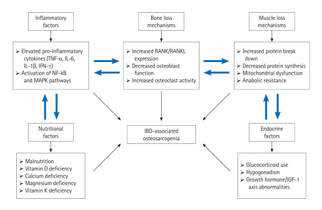
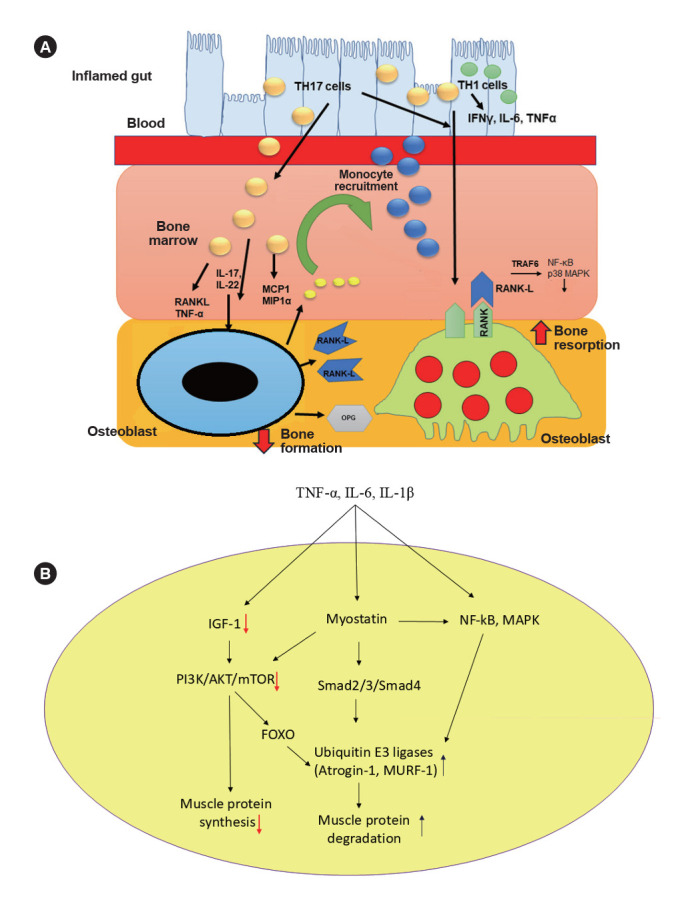
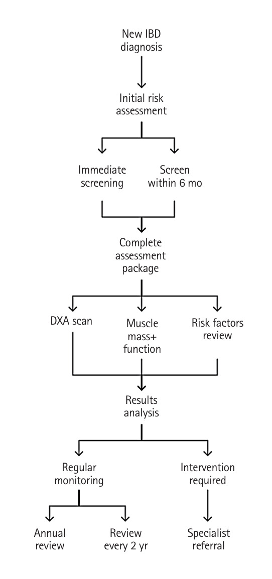
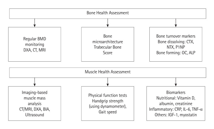
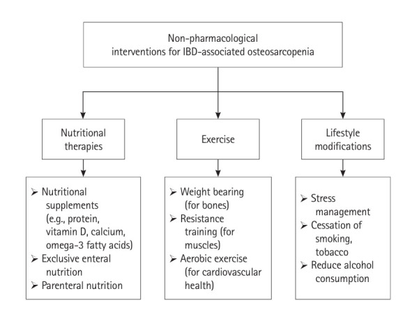
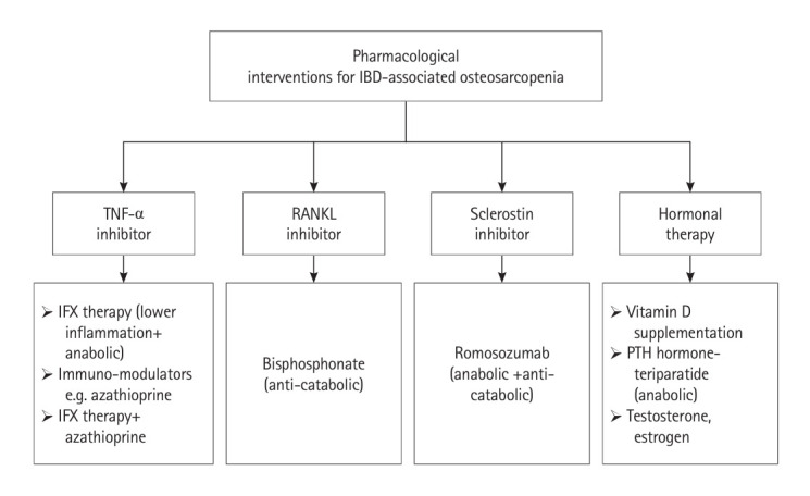
Inflammatory bowel disease (IBD) is a group of chronic inflammatory conditions affecting the gastrointestinal tract that can lead to multiple systemic complications. Among these, osteosarcopenia has emerged as a significant concern, characterized by the concurrent deterioration of bone density and muscle mass, strength, and function. This dual deterioration significantly elevates the risk of falls and fractures, thereby exacerbating morbidity and diminishing quality of life. The pathogenesis of IBD-associated osteosarcopenia is multifactorial, with chronic intestinal inflammation serving as a central driver. Pro-inflammatory cytokines simultaneously disrupt bone homeostasis and muscle metabolism, creating a catabolic environment that impacts both tissues. Nutritional deficiencies, common in IBD due to malabsorption and decreased dietary intake, further compromise both bone mineralization and muscle protein synthesis. Management requires a comprehensive approach combining nutritional optimization, structured physical therapy, and lifestyle modifications. Pharmacological interventions integrate diseasespecific treatments with targeted therapies including vitamin D supplementation, hormonal treatments, and bisphosphonates when indicated. This review synthesizes current evidence regarding the prevalence, pathogenesis, and clinical impact of osteosarcopenia in IBD, highlighting areas requiring further investigation.

Keywords: Bone loss; Inflammatory bowel disease; Muscle loss; Osteoporosis; Sarcopenia.


Conflict of interest statement

Conflict of Interest

No potential conflict of interest relevant to this article was reported.

Figures

Fig. 1.
Fig. 1.
Proposed pathophysiological mechanisms of IBD-associated osteosarcopenia. The diagram illustrates the interrelationships between inflammatory, nutritional, and endocrine factors contributing to both bone and muscle loss in IBD. Elevated pro-inflammatory cytokines activate pathways that disrupt bone homeostasis (via RANK/RANKL dysregulation) and muscle metabolism (via increased protein breakdown and mitochondrial dysfunction). These processes are further influenced by nutritional deficiencies and endocrine abnormalities, collectively resulting in IBD-associated osteosarcopenia. TNF, tumor necrosis factor; IL, interleukin; IFN, interferon; IGF, insulin-like growth factor; RANKL, receptor activator of nuclear factor kappa-β (RANK) ligand; IBD, inflammatory bowel disease.
Fig. 2.
Fig. 2.
(A) Schematic diagram illustrating RANKL-mediated bone loss in IBD. The inflamed gut in IBD shows increased presence of Th17 and Th1 cells, producing pro-inflammatory cytokines (IFN-γ, IL-6, TNF-α). Th17 cells migrate to and accumulate in the bone marrow and express RANKL and TNF-α, directly promoting osteoclast differentiation. Also, Th17 cells produce IL-17 and IL-22, stimulating local inf lammation and activating osteoblasts. Osteoblasts increase RANKL production, produce chemokines (MCP-1 and MIP-1α), which attract monocytes from the blood into the bone marrow. Osteoclasts are activated by RANKL binding to RANK receptors, resulting in increased bone resorption. Osteoblasts also produce OPG, which normally inhibits RANKL. However, in IBD, the balance is shifted towards increased RANKL activity. (B) Integrated signaling pathways in IBD-induced muscle wasting. This schematic diagram depicts the molecular mechanisms by which inflammatory cytokines (TNF-α, IL-6, IL-1β) promote muscle wasting through multiple pathways within muscle cells (represented by the yellow oval). The pathways include (1) decreased IGF-1 signaling leading to reduced PI3K/AKT/mTOR activity and enhanced FOXO signaling, resulting in decreased muscle protein synthesis; (2) increased myostatin expression activating Smad2/3/Smad4 signaling; and (3) NF-κB and MAPK activation. All these pathways converge to upregulate ubiquitin E3 ligases (Atrogin-1, MURF-1), leading to enhanced muscle protein degradation. Red downward arrows indicate decreased activity, while blue upward arrows indicate increased activity. This comprehensive pathway is particularly relevant in IBD patients receiving glucocorticoid therapy, as it illustrates how their treatment may potentially contribute to muscle wasting alongside inflammatory mediators. RANKL, receptor activator of nuclear factor kappa-β (RANK) ligand; IBD, inflammatory bowel disease; IFN, interferon; IL, interleukin; TNF, tumor necrosis factor; MCP-1, monocyte chemoattractant protein-1; MIP-1α, macrophage inflammatory protein-1α; OPG, osteoprotegerin; PI3K, phosphoinositide 3-kinase; AKT, protein kinase B; mTOR, mechanistic target of rapamycin; FOXO, forkhead box O; NF-κB, nuclear transcription factor-kappa B; MAPK, mitogen-activated protein kinase.
Fig. 3.
Fig. 3.
Diagnostic and monitoring algorithm for inflammatory bowel disease (IBD)-associated osteosarcopenia. The flowchart illustrates the systematic approach to screening, assessment, and monitoring of osteosarcopenia in newly diagnosed IBD patients. Following initial IBD diagnosis, patients undergo risk stratification to determine the timing of screening (immediate vs. within 6 months). A complete assessment package includes 3 core components: dual-energy X-ray absorptiometry (DXA) scan for bone health evaluation, muscle mass and function testing, and comprehensive risk factor review. Based on the results analysis, patients are directed to either regular monitoring (with annual or biennial reviews) or intervention pathways requiring specialist referral. This protocol ensures standardized evaluation and appropriate follow-up based on individual patient risk profiles and assessment outcomes.
Fig. 4.
Fig. 4.
Comprehensive assessment framework for bone and muscle health in IBD-associated osteosarcopenia. The diagram illustrates 2 major assessment domains: bone health (upper panel) and muscle health (lower panel). Bone health evaluation encompasses regular BMD monitoring through imaging techniques, bone microarchitecture analysis via trabecular bone scoring, and bone turnover markers for both resorption (CTX, NTX, P1NP) and formation (OC, ALP). Muscle health assessment includes imaging-based muscle mass quantification, functional testing of physical performance, and relevant biomarkers categorized as nutritional, inflammatory, and muscle-specific markers. IBD, inflammatory bowel disease; BMD, bone mineral density; DXA, dual-energy X-ray absorptiometry; CT, computed tomography; MRI, magnetic resonance imaging; CTX, C-terminal telopeptide of type I collagen; NTX, N-terminal telopeptide of type I collagen; P1NP; procollagen type 1 N propeptide; OC, osteocalcin; ALP, alkaline phosphatase; BIA, bioelectrical impedance analysis; CRP, C-reactive protein; IL, interleukin; TNF, tumor necrosis factor; IGF, insulin-like growth factor.
Fig. 5.
Fig. 5.
Non-pharmacological interventions for inflammatory bowel disease (IBD)-associated osteosarcopenia. Main categories of nonpharmacological interventions for managing osteosarcopenia in IBD patients are (1) nutritional therapies, comprising nutritional supplements (including protein, vitamin D, calcium, and omega-3 fatty acids), exclusive enteral nutrition, and parenteral nutrition; (2) exercise interventions, including weight-bearing exercises for bone health, resistance training for muscle strength, and aerobic exercise for cardiovascular fitness; and (3) lifestyle modifications, focusing on stress management, smoking/tobacco cessation, and reduction of alcohol consumption. These interventions collectively address bone health, muscle strength, and overall well-being in IBD patients with osteosarcopenia.
Fig. 6.
Fig. 6.
Pharmacological interventions for IBD-associated osteosarcopenia. Pharmacological interventions for IBD-associated osteosarcopenia. Major categories of pharmacological treatments for osteosarcopenia in IBD patients include: (1) TNF-α inhibitors, featuring IFX therapy (with anti-inflammatory and anabolic effects), immunomodulators like azathioprine, and combination therapy; (2) RANKL inhibitors, specifically bisphosphonate therapy with anti-catabolic properties; (3) sclerostin inhibitors, particularly romosozumab, which provides both anabolic and anti-catabolic effects; and (4) hormonal therapy options, including vitamin D supplementation, PTH hormoneteriparatide (anabolic), and sex hormones (testosterone and estrogen). IBD, inflammatory bowel disease; TNF, tumor necrosis factor; IFX, infliximab; RANKL, receptor activator of nuclear factor kappa-β ligand; PTH, parathyroid.

Similar articles

References

    1. Molodecky NA, Soon IS, Rabi DM, et al. Increasing incidence and prevalence of the inflammatory bowel diseases with time, based on systematic review. Gastroenterology. 2012;142:46–54. - PubMed
    1. Kaplan GG. The global burden of IBD: from 2015 to 2025. Nat Rev Gastroenterol Hepatol. 2015;12:720–727. - PubMed
    1. Sartor RB. Mechanisms of disease: pathogenesis of Crohn’s disease and ulcerative colitis. Nat Clin Pract Gastroenterol Hepatol. 2006;3:390–407. - PubMed
    1. Rogler G, Singh A, Kavanaugh A, Rubin DT. Extraintestinal manifestations of inflammatory bowel disease: current concepts, treatment, and implications for disease management. Gastroenterology. 2021;161:1118–1132. - PMC - PubMed
    1. Shen YH, Zhu H, Zhou L, et al. In inflammatory bowel disease and extraintestinal manifestations: what role does microbiome play? Eng Regen. 2023;4:337–348.

LinkOut - more resources