. 2025 Dec 1;15(4):459-470.
doi: 10.4103/mgr.MEDGASRES-D-24-00135. Epub 2025 Apr 29.

Magnesium-assisted hydrogen improves isoproterenol-induced heart failure

Affiliations

Magnesium-assisted hydrogen improves isoproterenol-induced heart failure

Fengbao Chen et al. Med Gas Res. .

Abstract

Heart failure (HF) is a leading cause of mortality among patients with cardiovascular disease and is often associated with myocardial apoptosis and endoplasmic reticulum stress (ERS). While hydrogen has demonstrated potential in reducing oxidative stress and ERS, recent evidence suggests that magnesium may aid in hydrogen release within the body, further enhancing these protective effects. This study aimed to investigate the cardioprotective effects of magnesium in reducing apoptosis and ERS through hydrogen release in a rat model of isoproterenol (ISO)-induced HF. Magnesium was administered orally to ISO-induced HF rats, which improved cardiac function, reduced myocardial fibrosis and cardiac hypertrophy, and lowered the plasma levels of creatine kinase-MB, cardiac troponin-I, and N-terminal B-type natriuretic peptide precursor in ISO-induced HF rats. It also inhibited cardiomyocyte apoptosis by upregulating B-cell lymphoma-2, downregulating Bcl-2-associated X protein, and suppressing ERS markers (glucose-related protein 78, activating transcription factor 4, and C/EBP-homologous protein). Magnesium also elevated hydrogen levels in blood, plasma, and cardiac tissue, as well as in artificial gastric juice and pure water, where hydrogen release lasted for at least four hours. Additionally, complementary in vitro experiments were conducted using H9C2 cardiomyocyte injury models, with hydrogen-rich culture medium as the intervention. Hydrogen-rich culture medium improved the survival and proliferation of ISO-treated H9C2 cells, reduced the cell surface area, inhibited apoptosis, and downregulated ERS pathway proteins. However, the protective effects of hydrogen were negated by tunicamycin (an inducer of ERS) in H9C2 cells. In conclusion, magnesium exerts significant cardioprotection by mitigating ERS and apoptosis through hydrogen release effects in ISO-induced HF.

Keywords: H9C2 cells; apoptosis; cardiac hypertrophy; endoplasmic reticulum stress; heart failure; hydrogen; isoproterenol; magnesium; myocardial fibrosis; rats.


Conflict of interest statement

Conflicts of interest: The authors declare no conflict of interest.

Figures

Figure 1
Figure 1
Flow chart of the animal and cell experiments. (A) Animal experiment flowchart. The purple area represents the time during which the magnesium, MgCl2, and HRW treatments were administered, and the blue area represents the period during which ISO subcutaneous injection induced HF. (B) Flowchart of the cell experiment. The purple area represents pretreatment with HRM for 24 hours, the blue area represents ISO treatment for 24 hours, and the yellow area represents treatment with tunicamycin for 12 hours. CCK-8: Cell Counting Kit-8; EdU: 5-ethynyl-2′-deoxyuridine; HRM: hydrogen-rich culture medium; HRW: hydrogen-rich saltwater; ISO: isoproterenol; MgCl2: magnesium chloride; Tm: tunicamycin; WGA: wheat germ agglutinin.
Figure 2
Figure 2
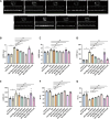
Magnesium ameliorates cardiac function in ISO-induced heart failure in rats. (A) Representative echocardiographic images. Rats in the ISO group exhibited cardiac dysfunction, whereas treatment with magnesium and HRW significantly improved cardiac function. (B–G) Changes in LVESD (B), LVEDD (C), LVESV (D), LVEDV (E), EF (F), and FS (G) from echocardiographic measurements. The data are expressed as the mean ± SEM (n = 5). **P < 0.01, vs. the control group; #P < 0.05, ##P < 0.01, vs. the ISO group (one-way analysis of variance followed by Tukey’s post hoc test). EF: Ejection fraction; FS: functional shortening; HRW: hydrogen-rich saltwater; ISO: isoproterenol; LVEDD: left ventricular end-diastolic diameter; LVESD: left ventricular end-systolic diameter; LVEDV: left ventricular end-diastolic volume; LVESV: left ventricular end-systolic volume; Mg: magnesium; MgCl2: magnesium chloride.
Figure 3
Figure 3
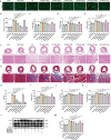
Magnesium alleviates myocardial hypertrophy and fibrosis in ISO-induced heart failure in rats. (A) Representative WGA staining of heart tissues. ISO group rats showed significant enlargement of myocardial cells, and magnesium and HRW effectively reduced the cell size. Scale bar: 20 μm. (B) Myocardial cell cross-sectional area. (C–E) Ratios of HW/BW (C), LVW/BW (D), and LVW/TL (E). (F) Representative HE staining of heart tissues (n = 3). Inflammatory cell infiltration and disordered arrangement of myocardial cells were observed in the heart tissue of ISO group rats, and magnesium and HRM alleviated this damage. Scale bar: 100 μm. (G) Representative Masson staining of heart tissues, where blue indicates collagen. Collagen deposition in the myocardium of ISO-treated rats significantly increased, but it was also reduced with magnesium and HRW treatments. Scale bar: 100 μm. (H) Quantification of myocardial fibrosis areas. (I–K) Plasma levels of CK-MB (I), cTn-I (J), and NT-proBNP (K). (L) Immunoblots of p-cTn-I and cTn-I proteins. (M) Quantification of p-cTn-I/cTn-I (n = 3). The data are expressed as the mean ± SEM (n = 5). **P < 0.01, vs. the control group; #P < 0.05, ##P < 0.01, vs. the ISO group (one-way analysis of variance followed by Tukey’s post hoc test). BW: Body weight; CK-MB: creatine kinase-MB; cTn-I: cardiac troponin-I; CVF: collagen volume fraction; GAPDH: glyceraldehyde-3-phosphate dehydrogenase; HE: hematoxylin-eosin; HRW: hydrogen-rich saltwater; HW: heart weight; ISO: isoproterenol; LVW: left ventricular weight; MCCA: myocardial cell cross-sectional area; Mg: magnesium; MgCl2: magnesium chloride; NT-proBNP: N-terminal B-type natriuretic peptide precursor; p-cTn-I: phosphorylated cardiac troponin-I; TL: tibial length; WGA: wheat germ agglutinin.
Figure 4
Figure 4
Magnesium suppresses apoptosis and ERS in ISO-induced heart failure in rats. (A) TUNEL staining (green) of heart tissues counterstained with DAPI (blue) (n = 5). The ISO group presented a significant increase in myocardial cell apoptosis, which was markedly reduced with magnesium and HRW treatments. Scale bar: 100 μm. (B) Percentage of TUNEL-positive cells. (C) Immunoblots of the Bax and Bcl-2 proteins. (D, E) Quantification of Bax (D) and Bcl-2 (E) expression. (F) Immunoblots of GRP78, ATF4, and CHOP. (G–I) Quantification of GRP78 (G), ATF4 (H), and CHOP (I) protein expression. The data are expressed as the mean ± SEM (n = 3). *P < 0.05, **P < 0.01, vs. the control group; #P < 0.05, ##P < 0.01, vs. the ISO group (one-way analysis of variance followed by Tukey’s post hoc test). ATF4: Activating transcription factor 4; Bax: Bcl-2 associated X protein; Bcl-2: B-cell lymphoma-2; CHOP: C/EBP-homologous protein; DAPI: 4’,6-diamidino-2-phenylindole; GAPDH: glyceraldehyde-3-phosphate dehydrogenase; GRP78: glucose-related protein78; HRW: hydrogen-rich saltwater; ISO: isoproterenol; Mg: magnesium; MgCl2: magnesium chloride; TUNEL: terminal dUTP nick end labeling.
Figure 5
Figure 5
Magnesium releases hydrogen in vitro and in vivo, which is correlated with heart failure parameters. (A, B) Hydrogen is released by magnesium in artificial gastric fluid and pure water (n = 3). (C–E) Hydrogen levels in rat blood (C), plasma (D), and heart tissues (E). (F) Plasma Mg2+ levels. The data are expressed as the mean ± SEM (n = 4–5). *P < 0.05, **P < 0.01, vs. the control group; #P < 0.05, ##P < 0.01, vs. the ISO group (two-way analysis or one-way analysis of variance followed by Tukey’s post hoc test). (G–I) Correlations of plasma hydrogen with EF (G), CVF (H), and MCCA (L). (J–L) Correlations of plasma Mg2+ levels with EF (J), CVF (K), and MCCA (L). CVF: Collagen volume fraction; EF: ejection fraction; HRW: hydrogen-rich saltwater; ISO: isoproterenol; MCCA: myocardial cell cross-sectional area; Mg: magnesium; MgCl2: magnesium chloride.
Figure 6
Figure 6
Hydrogen mitigates ISO-induced damage to H9C2 cardiomyocytes. (A–C) CCK-8 assay showing the effects of HRM and ISO on H9C2 cell viability. (D) EdU (green) and Hoechst (blue) staining of H9C2 cells (n = 6). The proliferation of cells in the ISO group was significantly reduced, whereas HRW treatment increased the proliferation of ISO-induced H9C2 cells. Scale bar: 100 μm. (E) Quantitative analysis of EdU incorporation. (F) WGA staining (green) with DAPI counterstaining (blue) (n = 3). The cells in the ISO group exhibited hypertrophy, while HRW treatment reduced their surface area. Scale bar: 20 μm. (G) Quantitative analysis of WGA staining. The data are expressed as the mean ± SEM. *P < 0.05, **P < 0.01, vs. the control group; #P < 0.05, ##P < 0.01, vs. the ISO group; @@P < 0.01, vs. the ISO + 150 μM HRM + tunicamycin group (one-way analysis of variance followed by Tukey’s post hoc test). CCK-8: Cell Counting Kit-8; EDU: 5-ethynyl-2′-deoxyuridine; HRM: hydrogen-rich culture medium; ISO: isoproterenol; Mg: magnesium; MgCl2: magnesium chloride; Tm: tunicamycin; WGA: wheat germ agglutinin.
Figure 7
Figure 7
Hydrogen inhibits ISO-induced apoptosis and ERS in H9C2 cells. (A) TUNEL staining of H9C2 cells. The apoptosis rate in the ISO group was significantly increased, whereas HRM treatment inhibited cell apoptosis. However, tunicamycin reversed the antiapoptotic effect of HRM. Scale bar: 50 μm. (B) Percentage of TUNEL-positive cells. (C) Immunoblots for Bax and Bcl-2 proteins. (D, E) Quantified levels of Bax (D) and Bcl-2 (E) proteins. (F) Immunoblots showing the protein levels of GRP78, ATF4, and CHOP. (G–I) Quantification of GRP78 (G), ATF4 (H), and CHOP (I) protein expression. The data are expressed as the mean ± SEM (n = 3). *P < 0.05, **P < 0.01, vs. the control group; #P < 0.05, ##P < 0.01, vs. the ISO group; @P < 0.05, @@P < 0.01, vs. the ISO + 150 μM HRM + tunicamycin group (one-way analysis of variance followed by Tukey’s post hoc test). ATF4: Activating transcription factor 4; Bax: Bcl-2 associated X protein; Bcl-2: B-cell lymphoma-2; CHOP: C/EBP-homologous protein; DAPI: 4′,6-diamidino-2-phenylindole; GAPDH: glyceraldehyde-3-phosphate dehydrogenase; GRP78: glucose-related protein 78; HRM: hydrogen-rich culture medium; ISO: isoproterenol; Mg: magnesium; MgCl2: magnesium chloride; Tm: tunicamycin; TUNEL: terminal dUTP nick end labeling.
Figure 8
Figure 8
The mechanism by which magnesium releases hydrogen to ameliorate heart failure. ATF4: Activating transcription factor4; Bax: Bcl-2 associated X protein; Bcl-2: B-cell lymphoma-2; CHOP: C/EBP-homologous protein; eif2α: eukaryotic translation initiation factor 2α; GRP78: glucose-related protein78; Mg: magnesium; PERK: protein kinase RNA-like endoplasmic reticulum kinase.

Similar articles

References

    1. Savarese G, Becher PM, Lund LH, Seferovic P, Rosano GMC, Coats AJS. Global burden of heart failure: a comprehensive and updated review of epidemiology. Cardiovasc Res. 2023;118:3272–3287. - PubMed
    1. Jiang H, Fang T, Cheng Z. Mechanism of heart failure after myocardial infarction. J Int Med Res. 2023;51:3000605231202573. - PMC - PubMed
    1. Cai Q, Li Y, Zhang Y, et al. Xinshubao tablet ameliorates myocardial injury against heart failure via the DCN/PPARα/PGC-1α/P300 pathway. Biomed Pharmacother. 2023;166:115285. - PubMed
    1. Wang Q, Liang S, Qian J, et al. OTUD1 promotes isoprenaline- and myocardial infarction-induced heart failure by targeting PDE5A in cardiomyocytes. Biochim Biophys Acta Mol Basis Dis. 2024;1870:167018. - PubMed
    1. Chouairi F, Levin A, Biegus J, Fudim M. Emerging devices for heart failure management. Prog Cardiovasc Dis. 2024;82:125–134. - PubMed

MeSH terms