Case Reports
. 2025 Jun 28;65(2):107-114.
doi: 10.3960/jslrt.24065. Epub 2025 Apr 30.

Composite mantle cell lymphoma and T-cell prolymphocytic leukemia: a case report

Affiliations
Case Reports

Composite mantle cell lymphoma and T-cell prolymphocytic leukemia: a case report

Emi Kemmoku et al. J Clin Exp Hematop. .

Abstract

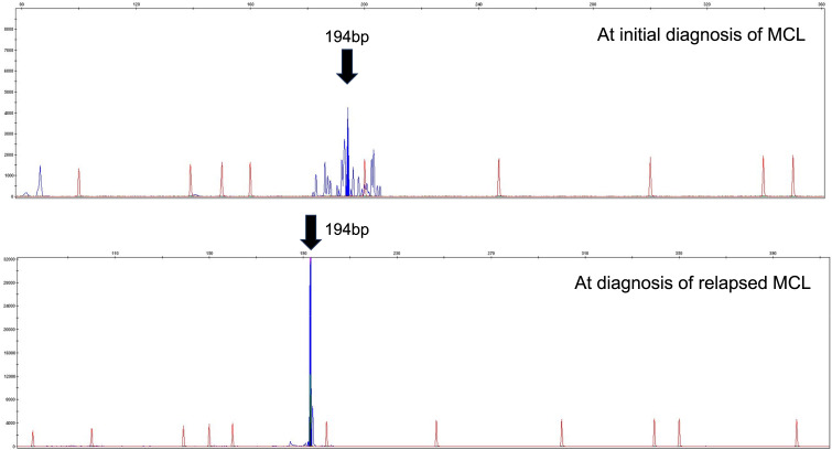
We encountered a patient with composite mantle cell lymphoma (MCL) and T-cell prolymphocytic leukemia (T-PLL) who presented with inactive disease to active T-PLL over 8 years. A 71-year-old man was diagnosed with MCL with an atypical T-cell population showing CD2+, CD3-, CD4+, CD7+, CD8-, and CD25+; however, the cause of the T-cell population could not be determined at the first MCL diagnosis. When MCL relapsed approximately 8 years after the initial treatment, T-PLL was definitively diagnosed using the T-PLL International Study Group criteria. MCL and T-PLL were determined to coexist in the lymph nodes and bone marrow by histological or flowcytometry analysis. Retrospective flow cytometry and T-cell receptor-polymerase chain reaction analysis of the stored samples suggested that the T-cell population noted at the time of initial MCL diagnosis eight years earlier was the same clone of T-PLL and the progression from inactive disease to active disease of his T-PLL. To the best of our knowledge, this is the first report of a composite MCL and T-PLL.

Keywords: T-cell prolymphocytic leukemia; composite lymphoma; from inactive to active disease; mantle cell lymphoma.


Conflict of interest statement

Shigeru Kusumoto received research funding from Kyowa Kirin Co., Ltd., Bristol-Myers Squibb, Abbvie, Meiji Seika Pharma, and honoraria from Chugai Pharmaceutical Co., Ltd., Daiichi Sankyo, Janssen Pharmaceutical K.K., and Ono Pharmaceutical Co., Ltd..

Toko Saito received research funding from Meiji Seika Pharma. Co., IQVIA, Abbvie, AstraZeneca, Nippon-shinyaku.

Kazuhito Yamamoto received research funding from AbbVie, Astra-Zeneca, Bayer, Bristol-Myers Squibb/Celgene, Chugai, Eisai, IQIVA/Genmab, IQIVA/Incyte, MSD, Mundipharma, Nippon Shinyaku, Novartis, Ono, Otsuka, Solasia Pharma, SymBio, Takeda, Yakult, and Zenyaku, and consulting fees from Meiji Seika Pharma, and honoraria from AbbVie, Astellas, Astra-Zeneca, Bristol-Myers Squibb/Celgene, Chugai, Daiichi Sankyo, Eisai, Eli Lilly, Genmab, Gilead, Incyte, IQIVA/HUYA, Janssen, Kyowa Kirin, Meiji Seika Pharma, Micron/Daiichi Sankyo, MSD, Mundipharma, Nippon Kayaku, Nippon Shinyaku, Novartis, Ono, Otsuka, Pfizer, Sanofi, Sumitomo Pharma, SymBio, Takeda, and Yakult.

Figures

Fig. 1
Fig. 1
Flowcytometry (FCM) analysis of the bone marrow at initial diagnosis of mantle cell lymphoma (MCL) Flowcytometry (FCM) analysis of the bone marrow at initial diagnosis of mantle cell lymphoma (MCL) The involvement of MCL cells (12% of mononuclear cells) showing CD5+, CD10-, CD19+, CD20+, light chain restriction to kappa are shown as the blue cells in the upper column. In addition, the involvement of the atypical T-cell population (9% of mononuclear cells) showing CD2+, CD3-, CD4+, CD7+, CD8-, CD25+, CCR4+ are shown as the pink cells in the lower column.
Fig. 2
Fig. 2
FCM analysis of the lymph node at diagnosis of relapsed MCL FCM analysis of the lymph node at diagnosis of relapsed MCL The involvement of MCL cells (87% of total cells) showing CD5+, CD10-, CD19+, CD20-, light chain restriction to kappa are shown as the blue cells in the upper column. In addition, the atypical T-cell population (7% of total cells) showing CD2+, CD3-, CD4+, CD7+, CD8-, CD25+) are shown as the pink cells in the lower column.
Fig. 3
Fig. 3
Peripheral blood smear This demonstrates the typical features of T-PLL. The tumor cells are medium-sized lymphoid cells with moderately condensed chromatin and a visible nucleolus (original magnification ×400).
Fig. 4
Fig. 4
Clinical course in a patient with composite MCL and T-PLL MCL, mantle cell lymphoma; T-PLL, T-cell prolymphocytic leukemia; PB, peripheral blood; BM, bone marrow; LN, lymph node; NA, not available; VR-CAP#8, 8 cycles of bortezimib, rituximab, cyclophosphamide, doxorubicin, and prednisone; R#12, 12 cycles of rituximab monotherapy; EBV-LPD, Epstein-Barr virus-associated B-cell lymphoproliferative disorder; R#8, 8 cycles of rituximab monotherapy; B#1, one cycle of bendamustine monotherapy; ALM, alemtuzumab; WBC, white blood cell; LDH, lactate dehydrogenase. Plus (+) indicates the involvement of MCL or T-PLL in the histological diagnosis or flow cytometry analysis.
Fig. 5
Fig. 5
Southern blot analysis of peripheral blood for T-cell receptor (TCR)-Cβ1 Southern blot analysis of peripheral blood for T-cell receptor (TCR)-Cβ1 Red arrows indicated the rearranged bands.
Fig. 6
Fig. 6
Bone marrow aspirate clot demonstrated that the involvement of mature T-cell neoplasm and relapsed mantle cell lymphoma Bone marrow aspirate clot demonstrated that the involvement of mature T-cell neoplasm and relapsed mantle cell lymphoma (original magnification ×40). HE, Hematoxylin and Eosin staining; CCND, CyclinD1 staining; FOXP3, Forkhead box P3 staining.
Fig. 7
Fig. 7
BIOMED-2 multiplex PCR assay for bone marrow (BM) samples at initial diagnosis of mantle cell lymphoma (MCL) and at diagnosis of relapsed MCL. It showed not only monoclonal rearrangements of the TCR gene, but also the same PCR product size in BM samples at the initial diagnosis of MCL and relapsed MCL.

Similar articles

References

    1. Campo E, Jaffe ES, Cook JR, et al. The International Consensus Classification of Mature Lymphoid Neoplasms: a report from the Clinical Advisory Committee. Blood. 2022; 140: 1229-1253. - PMC - PubMed
    1. Alaggio R, Amador C, Anagnostopoulos I, et al. The 5th edition of the World Health Organization Classification of Haematolymphoid Tumours: Lymphoid Neoplasms. Leukemia. 2022; 36: 1720-1748. - PMC - PubMed
    1. Raffeld M, Jaffe ES. bcl-1, t(11;14), and mantle cell-derived lymphomas. Blood. 1991; 78: 259-263. - PubMed
    1. Staber PB, Herling M, Bellido M, et al. Consensus criteria for diagnosis, staging, and treatment response assessment of T-cell prolymphocytic leukemia. Blood. 2019; 134: 1132-1143. - PMC - PubMed
    1. Garand R, Goasguen J, Brizard A, et al. Indolent course as a relatively frequent presentation in T-prolymphocytic leukaemia. Groupe Français d’Hématologie Cellulaire. Br J Haematol. 1998; 103: 488-494. - PubMed

Publication types